
Introduction
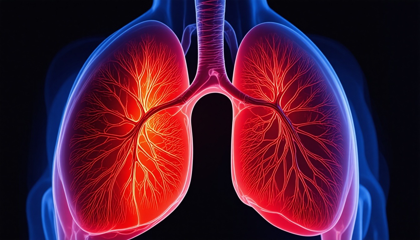
Breathing, or pulmonary ventilation, is the process by which air moves into and out of the lungs, enabling the exchange of oxygen and carbon dioxide between the atmosphere and the blood. This process is fundamental to life, providing the oxygen required for cellular respiration and removing the waste product carbon dioxide. The mechanism of breathing in humans is a complex interplay of muscles, bones, and pressure gradients, primarily driven by the diaphragm and intercostal muscles. Understanding the physical mechanism of gaseous exchange in humans requires a detailed exploration of the anatomy and physiology of the respiratory system, the mechanics of inhalation and exhalation, and the regulatory mechanisms that ensure efficient gas exchange. This article provides an in-depth, academic analysis of these processes, structured to facilitate comprehensive study.
The Mechanism of Gaseous Exchange in Humans: An Overview
Gaseous exchange in humans occurs at two main sites: the alveoli in the lungs (external respiration) and the tissues (internal respiration). The physical mechanism of breathing is essential for creating the airflow that brings fresh air to the alveoli and expels stale air. The process is governed by Boyle’s law, which states that at a constant temperature, the pressure of a gas is inversely proportional to its volume. By changing the volume of the thoracic cavity, the lungs are alternately inflated and deflated. This section introduces the key components involved in this mechanism.
- Thoracic Cavity: The chest cavity housing the lungs and heart, bounded by the rib cage, spine, and diaphragm.
- Pleural Cavities: The potential spaces between the visceral and parietal pleura, containing a thin layer of fluid that reduces friction and transmits forces.
- Intrapulmonary Pressure: The pressure inside the alveoli, which fluctuates during breathing.
- Intrapleural Pressure: The pressure within the pleural cavity, always negative relative to intrapulmonary pressure, keeping the lungs inflated.
These elements work together to create a pressure gradient that drives airflow. During inspiration, the thoracic cavity expands, decreasing intrapulmonary pressure below atmospheric pressure, causing air to rush in. During expiration, the thoracic cavity contracts, increasing intrapulmonary pressure above atmospheric, forcing air out. The following sections delve into the specific roles of the diaphragm and intercostal muscles.
Inhalation
Inhalation, or inspiration, is the active phase of breathing where air is drawn into the lungs. This process is primarily driven by the contraction of the diaphragm and the external intercostal muscles. The mechanism can be broken down into several detailed steps.
Diaphragmatic Contraction
The diaphragm is a dome-shaped skeletal muscle that separates the thoracic cavity from the abdominal cavity. When it contracts, it flattens and moves downward, increasing the vertical dimension of the thoracic cavity. This movement is the primary driver of inhalation, accounting for about 75% of the air intake during quiet breathing. The contraction is initiated by signals from the phrenic nerve (C3-C5). As the diaphragm descends, it pushes the abdominal organs downward, causing the abdomen to protrude slightly. This is why diaphragmatic breathing is also called abdominal breathing.
External Intercostal Muscle Action
The external intercostal muscles are located between the ribs, with fibers oriented obliquely downward and forward. When they contract, they elevate the ribs and sternum, increasing the anteroposterior and lateral dimensions of the thoracic cavity. This action is particularly important during deep breathing. The intercostal nerves (T1-T11) innervate these muscles. The combined effect of diaphragmatic and external intercostal contraction increases thoracic volume by up to 500-600 mL during quiet breathing, and much more during forced inhalation.
Pressure Changes During Inhalation
As the thoracic cavity expands, the volume of the pleural cavities increases, causing intrapleural pressure to become more negative (from about -5 cm H2O at rest to -8 cm H2O during quiet inspiration). This negative pressure is transmitted to the lungs, which are elastic and expand, increasing alveolar volume. According to Boyle’s law, the increased volume decreases alveolar pressure to about -1 cm H2O relative to atmospheric pressure. This pressure gradient drives air into the lungs until alveolar pressure equals atmospheric pressure at the end of inspiration.
Exhalation
Exhalation, or expiration, is the process by which air is expelled from the lungs. During quiet breathing, exhalation is a passive process, relying on the elastic recoil of the lungs and chest wall. but, during forced exhalation, it becomes an active process involving additional muscles.
Passive Exhalation
At the end of inhalation, the diaphragm and external intercostal muscles relax. The diaphragm returns to its dome shape, moving upward, and the ribs descend due to gravity and the elastic recoil of the costal cartilages. This reduces thoracic volume, increasing intrapleural pressure (to about -3 cm H2O) and alveolar pressure (to about +1 cm H2O). The positive pressure gradient forces air out of the lungs until alveolar pressure equals atmospheric pressure. The elastic recoil of the lungs is due to the presence of elastin fibers in the lung tissue and the surface tension of the alveolar fluid, which is reduced by surfactant.
Active Exhalation
During exercise or forced breathing, exhalation becomes active. The internal intercostal muscles, which have fibers oriented opposite to the external intercostals, contract to depress the ribs and sternum, reducing thoracic volume more rapidly. Additionally, the abdominal muscles (rectus abdominis, external and internal obliques, transversus abdominis) contract, increasing intra-abdominal pressure and pushing the diaphragm upward. This further decreases thoracic volume and increases alveolar pressure, allowing for more complete expulsion of air. The muscles of active exhalation are innervated by various spinal nerves.
Pressure Gradients in Exhalation
During quiet exhalation, the pressure changes are the reverse of inhalation. Alveolar pressure rises to about +1 cm H2O, creating a gradient of 1 cm H2O from the alveoli to the atmosphere. In forced exhalation, alveolar pressure can increase to +30 cm H2O or more, depending on effort. The intrapleural pressure also becomes less negative, but it remains negative throughout the breathing cycle to prevent lung collapse.
Role of the Diaphragm and Intercostal Muscles
The diaphragm and intercostal muscles are the primary muscles of respiration. Their coordinated action is essential for the mechanism of gaseous exchange in humans. This section provides a detailed analysis of their roles.
Diaphragm: The Primary Muscle of Inspiration
The diaphragm is a unique muscle in that it is both skeletal and involuntary in its basic rhythm, though it can be voluntarily controlled. It has three openings: the aortic hiatus, esophageal hiatus, and vena caval foramen. During contraction, the central tendon descends, increasing thoracic volume. The diaphragm’s contribution to inspiration is significant; even in quiet breathing, it accounts for the majority of air movement. In deep inspiration, the diaphragm can descend up to 10 cm. The phrenic nerve arises from the cervical plexus (C3-C5), and damage to this nerve can paralyze the diaphragm, leading to respiratory distress.
Intercostal Muscles: External and Internal
The external intercostal muscles are active during inspiration, while the internal intercostal muscles are primarily active during forced exhalation. The intercostal muscles are arranged in two layers: the external intercostals run obliquely downward and forward from the lower border of the rib above to the upper border of the rib below; the internal intercostals run obliquely downward and backward. The innermost intercostal muscles are a deeper layer that also aids in forced exhalation. Together, these muscles stabilize the chest wall and assist in changing thoracic volume. During inspiration, the external intercostals elevate the ribs, increasing the lateral and anteroposterior diameters of the chest. During forced exhalation, the internal intercostals depress the ribs.
Accessory Muscles of Respiration
also to the primary muscles, several accessory muscles assist during increased respiratory demand. These include the sternocleidomastoid, scalenes, pectoralis major and minor, serratus anterior, and latissimus dorsi for inspiration, and the abdominal muscles for exhalation. The sternocleidomastoid and scalenes elevate the first two ribs, while the pectoralis minor elevates the third to fifth ribs. During forced inspiration, these muscles are recruited to maximize thoracic expansion. During forced exhalation, the abdominal muscles are crucial for increasing intra-abdominal pressure.
Mechanics of Pulmonary Ventilation
Pulmonary ventilation is the total volume of air moved into and out of the lungs per minute. It is determined by the tidal volume (TV) and respiratory rate (RR). The mechanics involve the interplay of pressures, volumes, and resistances.
Lung Volumes and Capacities
Understanding lung volumes is essential for grasping the mechanism. Tidal volume (TV) is the volume of air inhaled or exhaled during a normal breath (about 500 mL). Inspiratory reserve volume (IRV) is the additional air that can be inhaled after a normal inhalation (about 3000 mL). Expiratory reserve volume (ERV) is the additional air that can be exhaled after a normal exhalation (about 1200 mL). Residual volume (RV) is the air remaining in the lungs after a forced exhalation (about 1200 mL). Lung capacities are sums of two or more volumes: vital capacity (VC = TV + IRV + ERV), inspiratory capacity (IC = TV + IRV), functional residual capacity (FRC = ERV + RV), and total lung capacity (TLC = VC + RV). These values vary with age, sex, body size, and health.
Compliance and Elasticity
Lung compliance is the measure of the lung’s ability to stretch and expand. It is defined as the change in lung volume per unit change in transpulmonary pressure (alveolar pressure minus intrapleural pressure). High compliance means the lungs are easily distensible, while low compliance indicates stiffness. Compliance is reduced in diseases like pulmonary fibrosis and increased in emphysema. Elasticity refers to the lung’s ability to recoil after being stretched. The elastic forces come from the elastin fibers in the lung tissue and the surface tension of the alveolar fluid. Surfactant, produced by type II alveolar cells, reduces surface tension, increasing compliance and preventing alveolar collapse.
Airway Resistance
Airflow through the airways is opposed by resistance, which is determined by the diameter of the airways and the viscosity of the gas. According to Poiseuille’s law, resistance is inversely proportional to the fourth power of the radius of the airway. so, even small changes in airway diameter (e.g., due to bronchoconstriction) can significantly affect airflow. The autonomic nervous system regulates bronchial smooth muscle tone: sympathetic stimulation causes bronchodilation, while parasympathetic stimulation causes bronchoconstriction. During breathing, airway resistance is minimal in the large airways but increases in the smaller bronchioles. but, the total cross-sectional area of the bronchioles is large, so overall resistance is low.
Regulation of Breathing
The rhythm of breathing is generated by the respiratory center in the medulla oblongata and pons. This center receives input from chemoreceptors, mechanoreceptors, and other factors to adjust the rate and depth of breathing according to the body’s needs.
Central and Peripheral Chemoreceptors
Central chemoreceptors are located in the medulla and are sensitive to changes in the pH of cerebrospinal fluid, which reflects arterial CO2 levels (via the blood-brain barrier). An increase in CO2 (hypercapnia) leads to a decrease in pH, stimulating the respiratory center to increase ventilation. Peripheral chemoreceptors are located in the carotid and aortic bodies and are sensitive to changes in arterial O2, CO2, and pH. Low O2 (hypoxemia) primarily stimulates the peripheral chemoreceptors, but this effect is weak unless O2 levels drop significantly. The response to CO2 is the most powerful driver of breathing under normal conditions.
Mechanoreceptors and Reflexes
Stretch receptors in the bronchial smooth muscle and lungs are activated by lung inflation. They send signals via the vagus nerve to inhibit inspiration, preventing overinflation (Hering-Breuer reflex). This reflex is more important in infants and during exercise. Irritant receptors in the airways respond to noxious stimuli, causing coughing and bronchoconstriction. J-receptors (juxtapulmonary capillary receptors) are stimulated by pulmonary congestion and edema, leading to rapid shallow breathing. These reflexes help protect the lungs and regulate breathing.
Voluntary Control and Other Factors
The cerebral cortex can voluntarily override the automatic respiratory center, allowing us to hold our breath or hyperventilate. but, this voluntary control is limited by chemical feedback. Other factors that influence breathing include body temperature (increased temperature increases rate), pain, emotion, and hormones like progesterone (which increases ventilation during pregnancy). The integration of these inputs ensures that breathing is precisely matched to metabolic demands.
Gas Exchange and Transport
Once air reaches the alveoli, oxygen diffuses into the blood and carbon dioxide diffuses out. This section details the process of gaseous exchange and how gases are transported in the blood.
Diffusion Across the Alveolar-Capillary Membrane
The alveolar-capillary membrane is extremely thin (0.5-1 μm) and has a large surface area (70-100 m²). Oxygen and carbon dioxide diffuse passively down their partial pressure gradients. The partial pressure of oxygen in the alveoli (PAO2) is about 100 mmHg, while in the pulmonary capillaries it is about 40 mmHg, so oxygen diffuses into the blood. Carbon dioxide has a partial pressure of about 45 mmHg in the capillaries and 40 mmHg in the alveoli, so it diffuses into the alveoli. The rate of diffusion is influenced by the thickness of the membrane, the surface area, and the solubility of the gases. Carbon dioxide is about 20 times more soluble than oxygen, so it diffuses more rapidly despite a smaller gradient.
Oxygen Transport
Oxygen is transported in the blood in two forms: dissolved (about 1.5%) and bound to hemoglobin (about 98.5%). Each hemoglobin molecule can bind up to four oxygen molecules, forming oxyhemoglobin. The binding is cooperative, meaning that the binding of one oxygen molecule increases the affinity for subsequent molecules. The oxygen-hemoglobin dissociation curve is sigmoidal, reflecting this cooperativity. Factors that shift the curve to the right (decreasing affinity) include increased temperature, decreased pH (Bohr effect), and increased 2,3-bisphosphoglycerate (2,3-BPG). These factors enhance oxygen unloading in tissues with high metabolic activity.
Carbon Dioxide Transport
Carbon dioxide is transported in three forms: dissolved (about 7%), as bicarbonate (about 70%), and bound to hemoglobin as carbaminohemoglobin (about 23%). In red blood cells, carbon dioxide reacts with water to form carbonic acid, catalyzed by carbonic anhydrase. Carbonic acid dissociates into bicarbonate and hydrogen ions. Bicarbonate is transported out of the red blood cell in exchange for chloride (chloride shift). The hydrogen ions are buffered by hemoglobin. In the lungs, these reactions reverse, releasing carbon dioxide for exhalation. The Haldane effect describes how deoxygenated hemoglobin has a greater capacity to carry carbon dioxide, facilitating its transport from tissues to lungs.
Factors Affecting Breathing Efficiency
Several factors can influence the efficiency of the physical mechanism of breathing, including posture, disease, and environmental conditions.
Posture and Gravity
In the upright position, gravity pulls the abdominal contents downward, allowing the diaphragm to descend more effectively. In the supine position, the abdominal contents push the diaphragm upward, reducing lung volume and compliance. This is why patients with respiratory distress often prefer to sit upright. Regional differences in ventilation and perfusion also occur due to gravity; in the upright lung, the bases are better ventilated and perfused than the apices.
Disease States
Obstructive lung diseases like asthma, chronic bronchitis, and emphysema increase airway resistance and reduce airflow. Restrictive lung diseases like pulmonary fibrosis reduce lung compliance, making it harder to expand the lungs. Neuromuscular disorders (e.g., muscular dystrophy, ALS) can weaken the respiratory muscles, impairing ventilation. These conditions alter the pressure-volume relationships and can lead to hypoventilation or respiratory failure.
Environmental Factors
High altitude reduces the partial pressure of oxygen in the atmosphere, leading to hypoxemia. The body adapts through hyperventilation, increased heart rate, and production of more red blood cells. Air pollution, including particulate matter and gases like ozone, can irritate the airways, causing bronchoconstriction and inflammation. Temperature and humidity also affect breathing; cold, dry air can cause bronchospasm in sensitive individuals.
Comparative Anatomy of Breathing
Comparing the human breathing mechanism with that of other animals provides evolutionary insights. for instance, birds have a unidirectional airflow system with air sacs, allowing for more efficient gas exchange during both inhalation and exhalation. Mammals, including humans, have a tidal flow system where air moves in and out through the same passages. Other animals like frogs use buccal pumping, and fish use gills with countercurrent exchange. The human diaphragm is a unique adaptation among mammals, but its basic function of creating negative pressure is common to all mammals.
Clinical Assessment of Breathing
Healthcare professionals assess breathing through various methods, including physical examination, spirometry, and imaging. This section outlines key clinical tools.
Spirometry
Spirometry measures lung volumes and capacities, including forced vital capacity (FVC) and forced expiratory volume in one second (FEV1). The FEV1/FVC ratio is used to differentiate obstructive from restrictive lung diseases. In obstructive diseases, the ratio is decreased (<0.7); in restrictive diseases, it is normal or increased. Spirometry is essential for diagnosing and monitoring respiratory conditions.
Blood Gas Analysis
Arterial blood gas (ABG) analysis measures partial pressures of oxygen (PaO2) and carbon dioxide (PaCO2), pH, and bicarbonate levels. It provides information about gas exchange efficiency and acid-base status. Hypoxemia (low PaO2) and hypercapnia (high PaCO2) indicate respiratory dysfunction. ABG is crucial in critical care settings.
Imaging
Chest X-rays and CT scans visualize the lungs, airways, and chest wall. They can detect pneumonia, tumors, pneumothorax, and other abnormalities. Dynamic imaging like fluoroscopy can assess diaphragm movement. These tools help diagnose structural causes of breathing impairment.
Breathing and Exercise
During exercise, the body’s demand for oxygen increases dramatically, and the breathing mechanism must adapt. This section explores the changes in ventilation during exercise.
Ventilatory Response to Exercise
At the onset of exercise, ventilation increases rapidly due to neural signals from the motor cortex and peripheral mechanoreceptors. As exercise continues, chemical stimuli (increased CO2, decreased pH, and later decreased O2) further drive ventilation. Tidal volume increases first, then respiratory rate. In moderate exercise, ventilation can increase from 6 L/min at rest to 50-100 L/min. In elite athletes, maximal ventilation can exceed 200 L/min.
Efficiency and Limitations
The breathing mechanism is highly efficient, but during intense exercise, the work of breathing can consume up to 10% of total oxygen consumption. The respiratory muscles may fatigue, limiting performance. Training can improve respiratory muscle endurance. Additionally, the oxygen-hemoglobin dissociation curve shifts right during exercise, enhancing oxygen unloading to muscles.
Breathing and Speech
Speech requires precise control of exhalation to produce sound. The physical mechanism of breathing must be modified to sustain phonation. During speech, exhalation is prolonged and controlled by the abdominal muscles and internal intercostals. The diaphragm plays a role in maintaining steady airflow. Respiratory patterns change: inspiratory phases are shorter and expiratory phases are longer. This coordination is learned and involves complex neural control.
Breathing and Sleep
During sleep, breathing patterns change, and the respiratory control system is less responsive. This can lead to sleep-disordered breathing like obstructive sleep apnea (OSA). In OSA, the upper airway collapses during sleep, causing intermittent hypoxia and arousals. The diaphragm and intercostal muscles continue to contract, but airflow is blocked. Treatment includes continuous positive airway pressure (CPAP) to keep the airway open. Understanding the physical mechanism of breathing is crucial for diagnosing and managing sleep-related breathing disorders.
Technological Advances in Respiratory Support
Modern medicine uses technology to assist or replace the breathing mechanism. This includes mechanical ventilators, which deliver positive pressure to inflate the lungs. Ventilators can be set to volume-controlled or pressure-controlled modes. Non-invasive ventilation (NIV) uses a mask to provide support without intubation. Extracorporeal membrane oxygenation (ECMO) bypasses the lungs entirely, oxygenating blood outside the body. These technologies are life-saving in critical care but require careful management to avoid complications like ventilator-induced lung injury.
Frequently Asked Questions
What is the primary muscle responsible for inhalation?
The primary muscle responsible for inhalation is the diaphragm. When it contracts, it flattens and moves downward, increasing the volume of the thoracic cavity and drawing air into the lungs. The external intercostal muscles also assist by elevating the ribs.
How does exhalation occur during quiet breathing?
During quiet breathing, exhalation is a passive process. The diaphragm and external intercostal muscles relax, allowing the elastic recoil of the lungs and chest wall to decrease thoracic volume. This increases alveolar pressure above atmospheric pressure, pushing air out of the lungs.
What is the role of the intercostal muscles in forced exhalation?
During forced exhalation, the internal intercostal muscles contract to depress the ribs and sternum, reducing thoracic volume. Additionally, the abdominal muscles contract to push the diaphragm upward, further decreasing volume and increasing pressure.
How does the diaphragm contribute to the mechanism of gaseous exchange?
The diaphragm creates the pressure gradient that drives airflow. Its contraction expands the thoracic cavity, lowering intrapulmonary pressure and drawing air into the lungs. This brings fresh oxygen to the alveoli for diffusion into the blood and removes carbon dioxide from the alveoli.
What factors can affect the efficiency of breathing?
Factors include posture, lung compliance, airway resistance, disease states (e.g., asthma, fibrosis), neuromuscular function, and environmental conditions like altitude and pollution. These can alter the pressure-volume relationships and impede gas exchange.
How is breathing regulated by the nervous system?
The respiratory center in the medulla oblongata generates the basic rhythm of breathing. It receives input from central and peripheral chemoreceptors that monitor CO2, O2, and pH, as well as from mechanoreceptors in the lungs and airways. This input adjusts the rate and depth of breathing to meet metabolic demands.
What is the difference between external and internal respiration?
External respiration refers to the exchange of gases between the alveoli and the pulmonary capillaries, where oxygen enters the blood and carbon dioxide leaves. Internal respiration is the exchange of gases between the systemic capillaries and the tissues, where oxygen is delivered to cells and carbon dioxide is picked up.
Can the diaphragm be voluntarily controlled?
Yes, the diaphragm is under both involuntary and voluntary control. The automatic rhythm is generated by the respiratory center, but the cerebral cortex can override it, allowing us to voluntarily hold our breath, breathe deeply, or speak. but, voluntary control is limited by chemical feedback.
What happens to breathing during exercise?
During exercise, ventilation increases to meet the higher oxygen demand and remove excess carbon dioxide. Tidal volume and respiratory rate increase. Neural and chemical stimuli drive this response. The respiratory muscles work harder, and in intense exercise, the work of breathing can become a limiting factor.
How does sleep affect breathing?
During sleep, the respiratory center is less responsive to CO2 and O2 changes, and muscle tone decreases, which can narrow the upper airway. This can lead to sleep-disordered breathing like obstructive sleep apnea, characterized by repeated pauses in breathing due to airway collapse.
Conclusion
The physical mechanism of breathing in humans is a marvel of biological engineering, integrating muscular action, pressure gradients, and neural control to achieve efficient gaseous exchange. The diaphragm and intercostal muscles play central roles in inhalation and exhalation, with the diaphragm being the primary driver of inspiration. Understanding the mechanism of gaseous exchange in humans requires a deep appreciation of the anatomy, physics, and regulation of breathing. From the passive elastic recoil during quiet exhalation to the active recruitment of accessory muscles during forced breathing, every aspect is finely tuned to meet the body’s metabolic needs. Clinical assessment tools like spirometry and blood gas analysis allow us to evaluate this mechanism, and technological advances provide support when it fails. This comprehensive guide has covered the essential topics for academic study, emphasizing the key roles of the diaphragm, intercostal muscles, and the pressure gradients that drive airflow. Mastery of these concepts is fundamental for students of medicine, physiology, and related fields.
Embryological Development of the Respiratory Muscles and Thoracic Cavity
The physical mechanism of breathing is established during embryonic development, with the diaphragm and intercostal muscles originating from distinct mesodermal sources. The diaphragm develops from four embryonic structures: the septum transversum (central tendon), pleuroperitoneal membranes (posterolateral portions), dorsal mesentery of the esophagus (crura), and body wall musculature (peripheral muscular portion). By the eighth week of gestation, the diaphragm is fully formed, separating the thoracic and abdominal cavities. The phrenic nerve, derived from cervical somites C3-C5, innervates the diaphragm, reflecting its cervical origin. The intercostal muscles develop from the thoracic somites, with myoblasts migrating to form the external, internal, and innermost layers. The rib cage forms from the costal processes of the thoracic vertebrae, providing attachment points for these muscles. Understanding this development is crucial for comprehending congenital anomalies such as congenital diaphragmatic hernia, where abdominal organs herniate into the chest, impairing lung development and breathing mechanics. Such conditions highlight the importance of structural integrity for normal ventilation.
Biomechanics of the Rib Cage and Spine
The rib cage and spine form a semi-rigid but flexible framework that moves during breathing. The ribs articulate with the thoracic vertebrae posteriorly and with the sternum anteriorly via costal cartilages. The ribs are angled downward and forward, and their movement during inspiration is often described as a ‘bucket handle’ motion (elevation of the ribs increases the lateral diameter of the chest) and a ‘pump handle’ motion (elevation of the sternum increases the anteroposterior diameter). The external intercostal muscles, by contracting, pull the ribs upward and outward. The spine also plays a role: thoracic extension (straightening) can increase thoracic volume, while flexion reduces it. In conditions like kyphosis (excessive curvature of the thoracic spine), lung expansion may be limited, reducing vital capacity. The costovertebral and costotransverse joints allow the ribs to rotate and glide, facilitating smooth movement. The biomechanics of the chest wall are essential for efficient ventilation, and any restriction, such as in ankylosing spondylitis where the costovertebral joints fuse, can severely impair breathing.
Pleural Pressure Dynamics and Lung-Pleura Coupling
The pleural cavity is a potential space filled with a thin layer of pleural fluid, which couples the lungs to the chest wall. The intrapleural pressure is normally negative (about -5 cm H2O at rest) due to the opposing elastic recoil of the lungs (tending to collapse) and the chest wall (tending to expand). This negative pressure keeps the lungs inflated and allows them to follow chest wall movements. During inspiration, the chest wall expands, and the intrapleural pressure becomes more negative (e.g., -8 cm H2O), which expands the lungs. If the pleural space is breached (e.g., pneumothorax), the intrapleural pressure equalizes with atmospheric pressure, and the lung collapses. The pleural fluid also acts as a lubricant, reducing friction during breathing. The dynamics of pleural pressure are critical for understanding conditions like pleural effusion, where fluid accumulation increases intrapleural pressure and compresses the lung, reducing compliance and causing dyspnea.
Work of Breathing and Respiratory Muscle Energetics
The work of breathing is the energy expended by the respiratory muscles to overcome elastic forces (lung and chest wall compliance), resistive forces (airway resistance), and inertial forces (tissue movement). At rest, the work of breathing is about 0.5-1% of total body oxygen consumption, but during exercise, it can increase to 10% or more. The work is calculated as the area under the pressure-volume curve. In healthy individuals, most of the work is done during inspiration (active), while expiration is passive. In obstructive lung diseases, airway resistance increases, requiring more work to overcome resistive forces. In restrictive diseases, elastic work increases. Respiratory muscle fatigue can occur when the energy demand exceeds supply, leading to hypoventilation and respiratory failure. The diaphragm, being the primary muscle, is highly oxidative and rich in mitochondria, but it can fatigue under high loads. Training the respiratory muscles can improve endurance, which is beneficial for patients with chronic respiratory disease and athletes.
Effects of Age on Breathing Mechanics
Aging affects the physical mechanism of breathing in several ways. Lung elasticity decreases due to loss of elastin, leading to increased compliance and reduced elastic recoil. This results in a higher residual volume and lower vital capacity. The chest wall becomes stiffer due to calcification of costal cartilages and arthritis of costovertebral joints, decreasing compliance. Respiratory muscle strength declines, with the diaphragm losing about 25% of its mass by age 70. These changes reduce the efficiency of ventilation, making older adults more susceptible to respiratory infections and hypoxemia. The response to hypoxia and hypercapnia is blunted, increasing the risk of respiratory failure during illness. Understanding age-related changes is important for geriatric medicine and for interpreting pulmonary function tests in the elderly.
Neuromuscular Control of the Respiratory Pump
The respiratory pump is controlled by a complex neural network involving the brainstem, spinal cord, and peripheral nerves. The central pattern generator for breathing is located in the pre-Bötzinger complex in the medulla, which generates inspiratory rhythm. This rhythm is modulated by inputs from the pons (pneumotaxic center, apneustic center) and chemoreceptors. The phrenic nerve and intercostal nerves transmit motor commands to the diaphragm and intercostal muscles. Sensory feedback from muscle spindles and tendon organs in the respiratory muscles, as well as from lung stretch receptors, fine-tunes the motor output. Damage to the spinal cord above C3-C5 (e.g., in spinal cord injury) can paralyze the diaphragm, requiring mechanical ventilation. Conditions like amyotrophic lateral sclerosis (ALS) progressively weaken respiratory muscles, leading to respiratory failure. Understanding this control system is essential for developing treatments for central sleep apnea, where the brainstem fails to generate adequate respiratory drive.
Breathing and Cardiovascular Interaction
Breathing affects cardiovascular function through changes in intrathoracic pressure. During inspiration, the negative intrathoracic pressure increases venous return to the right heart (by increasing the pressure gradient from the systemic veins to the right atrium), which increases right ventricular stroke volume. but, it also increases pulmonary vascular resistance, slightly decreasing left ventricular filling. This leads to a small decrease in systolic blood pressure during inspiration (pulsus paradoxus), which is exaggerated in conditions like cardiac tamponade or severe asthma. During expiration, the opposite occurs. The respiratory cycle also influences heart rate variability (respiratory sinus arrhythmia), where heart rate increases during inspiration and decreases during expiration, mediated by vagal tone. This interaction is important in critical care, where positive pressure ventilation can reduce venous return and cardiac output. Understanding cardiopulmonary interaction is vital for managing patients on mechanical ventilation.
Breathing in Extreme Environments: Diving and High Altitude
In diving, the physical mechanism of breathing is challenged by increased ambient pressure. As depth increases, the partial pressure of gases increases, and the work of breathing increases due to higher gas density. The diaphragm and intercostal muscles must work harder to overcome the increased resistance. At great depths, breathing compressed air can lead to nitrogen narcosis, while breathing pure oxygen can cause oxygen toxicity. The pressure-volume relationship of the lungs is also affected; the lungs are compressible, but excessive pressure can cause barotrauma. In high altitude, the low partial pressure of oxygen triggers hyperventilation, which can lead to respiratory alkalosis. The body adapts through increased ventilation, but this is limited by the work of breathing and the risk of respiratory muscle fatigue. Acclimatization involves increased erythropoietin production and changes in oxygen-hemoglobin affinity. These extreme environments test the limits of the breathing mechanism and provide insights into its adaptability.
Breathing and Acid-Base Balance
The respiratory system plays a crucial role in maintaining acid-base balance by regulating the partial pressure of carbon dioxide (PaCO2). Carbon dioxide, when dissolved in blood, forms carbonic acid, which dissociates to bicarbonate and hydrogen ions. Thus, changes in PaCO2 directly affect blood pH. Hyperventilation (increased breathing) lowers PaCO2, causing respiratory alkalosis, while hypoventilation (decreased breathing) raises PaCO2, causing respiratory acidosis. The respiratory center responds to changes in pH and PaCO2 to adjust ventilation. In metabolic acidosis (e.g., diabetic ketoacidosis), the respiratory center is stimulated to increase ventilation (Kussmaul breathing) to blow off CO2 and compensate. In metabolic alkalosis, ventilation is decreased to retain CO2. The respiratory compensation is rapid, occurring within minutes, and is essential for maintaining pH within a narrow range. Understanding this relationship is critical for interpreting blood gas results and managing patients with acid-base disorders.
Respiratory Muscle Training and Rehabilitation
Respiratory muscle training (RMT) involves exercises to strengthen the diaphragm and intercostal muscles, often using devices that provide resistance during inspiration or expiration. RMT can improve respiratory muscle strength and endurance, reduce dyspnea, and enhance exercise capacity in patients with chronic obstructive pulmonary disease (COPD), heart failure, and neuromuscular disorders. Techniques include inspiratory muscle training (IMT) using a threshold loading device, and expiratory muscle training (EMT) for cough effectiveness. In athletes, RMT can improve ventilatory efficiency and delay respiratory muscle fatigue. The mechanisms include increased muscle fiber hypertrophy, improved oxidative capacity, and neural adaptations. but, the evidence for RMT in improving clinical outcomes is mixed, and it isn’t universally recommended. Nonetheless, understanding the physiological basis of RMT helps in designing rehabilitation programs for respiratory patients.
Breathing and Voice Production
Voice production requires precise control of exhalation to generate a steady airflow through the vocal cords. The diaphragm and abdominal muscles work together to maintain a constant subglottic pressure (around 5-10 cm H2O) during phonation. The intercostal muscles regulate the rate of chest wall collapse. During speech, inspiratory phases are short and deep, while expiratory phases are prolonged and controlled. Professional singers and speakers train to optimize this coordination, using diaphragmatic breathing to support the voice. Pathologies like vocal cord paralysis or muscle tension dysphonia can disrupt this mechanism, leading to voice disorders. Respiratory therapy can help improve breath support for voice production. The integration of breathing and phonation is a complex motor skill that highlights the versatility of the respiratory system.
Breathing and Emotional States
Emotions like anxiety, fear, and excitement can alter breathing patterns. The limbic system and hypothalamus influence the respiratory center, leading to rapid, shallow breathing during panic attacks (hyperventilation), or sighing during sadness. Stress can lead to chronic hyperventilation, causing symptoms like dizziness, paresthesias, and chest tightness. Conversely, slow, deep breathing techniques (e.g., pranayama in yoga) can activate the parasympathetic nervous system, reducing heart rate and blood pressure. This is the basis for breathing exercises in stress management and meditation. The interaction between emotion and breathing is bidirectional; altering breathing can affect emotional states. Understanding this connection is important for treating conditions like anxiety disorders and for promoting relaxation techniques.
Breathing and the Lymphatic System
The lymphatic system relies on movement of the thoracic cavity to propel lymph through the thoracic duct and right lymphatic duct. The negative pressure during inspiration and the compression of lymphatic vessels by respiratory muscles and the diaphragm facilitate lymph flow. The thoracic duct, which drains lymph from most of the body, passes through the diaphragm and is compressed during inspiration, pushing lymph forward. This mechanism is important for immune function and fluid balance. In conditions like lymphedema, impaired lymph flow can be exacerbated by reduced respiratory movement. Deep breathing exercises are sometimes recommended to promote lymphatic drainage. The connection between breathing and lymphatic circulation is an often-overlooked aspect of respiratory physiology.
Breathing and the Gastrointestinal System
The diaphragm and abdominal muscles interact with the gastrointestinal (GI) system. During inspiration, the descending diaphragm increases intra-abdominal pressure, which can compress the stomach and intestines, aiding in digestion and preventing gastroesophageal reflux? Actually, increased intra-abdominal pressure can promote reflux, which is why some people experience heartburn when breathing deeply after a meal. The diaphragm also plays a role in the mechanism of vomiting, where a deep inspiration is followed by contraction of the abdominal muscles to increase pressure and expel stomach contents. also, the rhythmic movement of the diaphragm massages the abdominal organs, potentially stimulating peristalsis. This interaction is complex and not fully understood, but it highlights the multi-system effects of the breathing mechanism.
Breathing and the Immune System
The respiratory system is a major interface with the environment and is constantly exposed to pathogens. The physical mechanism of breathing helps to clear inhaled particles and microbes through mucociliary clearance and cough. The diaphragm and intercostal muscles generate the forceful exhalation needed for an effective cough. Additionally, breathing patterns can influence immune function; for instance, hyperventilation can lead to hypocapnia, which may alter immune cell function. Chronic stress and altered breathing patterns are associated with increased susceptibility to infections. Conversely, deep breathing exercises may enhance immune function by reducing stress hormones. The relationship between breathing and immunity is an emerging area of research with potential clinical applications.
Breathing and the Renal System
The kidneys and lungs work together to maintain acid-base balance. The respiratory system provides rapid compensation for acid-base disturbances, while the kidneys provide slower, long-term compensation. for instance, in chronic respiratory acidosis (e.g., from COPD), the kidneys retain bicarbonate to normalize pH. In chronic respiratory alkalosis, the kidneys excrete bicarbonate. The physical mechanism of breathing itself doesn’t directly affect renal function, but the changes in blood gases and pH influence renal blood flow and glomerular filtration rate. Hypoxemia can cause renal vasoconstriction, reducing urine output. Understanding this interplay is important for managing critically ill patients with multi-organ dysfunction.
Breathing and the Endocrine System
Several hormones affect breathing, and breathing can affect hormone release. Progesterone, for instance, stimulates ventilation, which is why pregnant women often experience increased breathing rate. Thyroid hormones increase metabolic rate and thus ventilation. Cortisol can affect respiratory muscle function. Conversely, hyperventilation can cause changes in blood pH that affect hormone secretion. The stress response involves increased ventilation, which may help prepare the body for fight or flight. The endocrine system modulates the respiratory control system, and disruptions can lead to breathing disorders. For instance, hypothyroidism can lead to hypoventilation and sleep apnea.
Breathing and Pharmacology
Many drugs affect the physical mechanism of breathing. Opioids depress the respiratory center, reducing ventilatory drive and leading to hypoventilation. Benzodiazepines and barbiturates have similar effects. Bronchodilators like beta-agonists relax airway smooth muscle, reducing resistance and improving airflow. Corticosteroids reduce airway inflammation. Respiratory stimulants like caffeine and theophylline can increase ventilatory drive. Understanding how drugs affect breathing is crucial for safe prescribing, especially in patients with respiratory compromise. The mechanism of action and side effects must be considered to avoid respiratory depression or other adverse effects.
Breathing and Critical Care: Mechanical Ventilation Strategies
In critical care, mechanical ventilation supports or replaces the physical mechanism of breathing. Different modes of ventilation are used depending on the patient’s condition. Volume-controlled ventilation delivers a set tidal volume, while pressure-controlled ventilation delivers a set inspiratory pressure. Positive end-expiratory pressure (PEEP) is used to keep alveoli open and improve oxygenation. Protective lung ventilation strategies, such as low tidal volumes and limited plateau pressures, are used to prevent ventilator-induced lung injury in patients with acute respiratory distress syndrome (ARDS). Understanding the pressure-volume curves and lung mechanics is essential for setting ventilators. The role of the diaphragm and intercostal muscles is often suppressed by sedation and paralysis, but spontaneous breathing efforts can be allowed in certain modes to prevent muscle atrophy. Weaning from ventilation requires assessing the patient’s ability to resume spontaneous breathing.
Breathing and Surgical Considerations
During surgery, anesthesia affects the physical mechanism of breathing. General anesthetics depress the respiratory center and reduce muscle tone, including the diaphragm and intercostals. Mechanical ventilation is often required. Positioning during surgery (e.g., prone, lateral) can affect lung mechanics and ventilation-perfusion matching. Postoperative pulmonary complications like atelectasis and pneumonia are common, and preventive measures include incentive spirometry, deep breathing exercises, and early mobilization. The diaphragm can be affected by upper abdominal or thoracic surgery, leading to diaphragmatic dysfunction. Understanding the effects of surgery on breathing is important for anesthesiologists and surgeons to optimize patient outcomes.
Breathing and Sleep Disorders: Beyond Apnea
Obstructive sleep apnea (OSA) is the most common sleep-disordered breathing, but other conditions like central sleep apnea (CSA), Cheyne-Stokes respiration, and hypoventilation syndromes also affect breathing during sleep. In OSA, the pharyngeal airway collapses, requiring increased respiratory effort to overcome obstruction, leading to arousals. In CSA, the respiratory center fails to generate adequate drive. Cheyne-Stokes respiration is characterized by a crescendo-decrescendo pattern of breathing, often seen in heart failure. Treatment includes CPAP, adaptive servo-ventilation, and oxygen therapy. The physical mechanism of breathing during sleep is influenced by changes in muscle tone, chemoresponsiveness, and sleep stage. Understanding these disorders is crucial for sleep medicine.
Breathing and Respiratory Rehabilitation
Pulmonary rehabilitation is a comprehensive program that includes exercise training, education, and breathing techniques for patients with chronic respiratory disease. Techniques like pursed-lip breathing and diaphragmatic breathing help improve ventilation efficiency, reduce dyspnea, and enhance quality of life. Pursed-lip breathing creates backpressure in the airways, preventing collapse and prolonging exhalation. Diaphragmatic breathing encourages the use of the diaphragm rather than accessory muscles. These techniques are taught to patients with COPD, asthma, and other conditions. The evidence supports their effectiveness in improving exercise tolerance and reducing symptoms. Rehabilitation also includes inspiratory muscle training as discussed earlier.
Breathing and Palliative Care
In palliative care, managing dyspnea is a priority. The physical mechanism of breathing may be compromised by advanced disease, such as cancer, heart failure, or neurological conditions. Opioids are used to reduce the sensation of breathlessness by depressing the respiratory center and reducing anxiety. Oxygen therapy, fan therapy, and positioning can help. Breathing techniques like relaxation and guided imagery can also provide relief. Understanding the pathophysiology of dyspnea, which involves both the physical work of breathing and central perception, is important for effective palliation.
Future Directions in Respiratory Physiology Research
Research continues to uncover new aspects of the physical mechanism of breathing. Advances in imaging (e.g., dynamic MRI, ultrasound) allow real-time visualization of diaphragm and chest wall movement. Computational modeling of lung mechanics helps predict disease progression and optimize ventilation strategies. Stem cell research aims to regenerate damaged respiratory muscles. Neuroprosthetics may restore breathing in spinal cord injury. The role of the microbiome in lung health and breathing is being explored. These future directions promise to improve our understanding and treatment of respiratory disorders.
Conclusion
The physical mechanism of breathing in humans is a multifaceted process involving the coordinated action of the diaphragm, intercostal muscles, rib cage, and neural control systems. This comprehensive guide has explored the intricate details of inhalation and exhalation, the role of the diaphragm and intercostal muscles, the mechanism of gaseous exchange, and the numerous factors that influence breathing efficiency. From embryological development to critical care applications, the depth of this topic reflects its fundamental importance in human physiology. Understanding these mechanisms is essential for healthcare professionals, researchers, and students alike, as it underpins the diagnosis and management of respiratory disorders and informs therapeutic interventions. The continued exploration of breathing mechanics will undoubtedly yield new insights and improve patient care.
{
“content”: “
Breathing and the Autonomic Nervous System
The autonomic nervous system (ANS) plays a crucial role in modulating the physical mechanism of breathing, primarily through the sympathetic and parasympathetic divisions. The parasympathetic system, via the vagus nerve, promotes bronchoconstriction and increases mucus secretion, which can increase airway resistance. It also influences the respiratory center in the medulla, contributing to the fine-tuning of breathing rate and depth. In contrast, the sympathetic system, through the release of norepinephrine and epinephrine, causes bronchodilation, reducing airway resistance and improving airflow. This is particularly important during exercise or stress, when increased ventilation is needed. The balance between these two systems is crucial for maintaining airway patency and optimal gas exchange. Dysregulation of the ANS can contribute to respiratory disorders such as asthma, where exaggerated parasympathetic tone leads to bronchoconstriction, or in chronic obstructive pulmonary disease (COPD), where sympathetic overactivity may contribute to increased work of breathing. Understanding this autonomic regulation is essential for developing pharmacological interventions that target specific receptors (e.g., beta-2 agonists for bronchodilation, anticholinergics for reducing parasympathetic effects).
Breathing and the Immune System: Defense Mechanisms
The respiratory system is constantly exposed to pathogens and particulate matter, and the physical mechanism of breathing is integral to immune defense. The mucociliary escalator, composed of ciliated epithelial cells and mucus, traps inhaled particles and moves them upward to be swallowed or expectorated. The effectiveness of this clearance depends on the depth and frequency of breathing; shallow breathing can reduce clearance, increasing infection risk. Coughing and sneezing are forceful expiratory maneuvers that expel irritants and pathogens from the airways. These reflexes involve a deep inspiration followed by a powerful contraction of the diaphragm, intercostal muscles, and abdominal muscles, generating high airflow velocities. The respiratory muscles also play a role in the production of surfactant, which contains immune components like surfactant proteins A and D that opsonize pathogens and enhance phagocytosis. Chronic respiratory conditions like COPD are associated with impaired mucociliary clearance and increased susceptibility to infections, highlighting the importance of the breathing mechanism in immune function.
Breathing and the Musculoskeletal System: Posture and Alignment
Posture significantly influences the mechanical efficiency of breathing. An upright posture allows the diaphragm to descend fully, maximizing thoracic expansion. Slouching or forward head posture compresses the thoracic cavity, restricting diaphragm movement and reducing lung volumes. The intercostal muscles and accessory muscles must work harder to compensate, increasing the work of breathing. Over time, poor posture can lead to muscle imbalances, such as tight pectorals and weak upper back muscles, further impairing breathing. Conversely, deep breathing exercises can improve posture by strengthening the diaphragm and core muscles. In conditions like scoliosis, the curvature of the spine distorts the rib cage, impairing lung expansion and leading to restrictive lung disease. Physical therapy focusing on postural correction and respiratory muscle training can improve breathing mechanics in these patients. The relationship between posture and breathing is bidirectional, and optimizing posture is a key component of respiratory rehabilitation.
Breathing and the Nervous System: Pain and Breathing
Pain, particularly in the chest, abdomen, or back, can significantly alter the physical mechanism of breathing. Acute pain often leads to shallow, rapid breathing (tachypnea) as the patient splints the painful area to minimize movement. This reduces tidal volume and can lead to atelectasis (collapse of alveoli) and hypoxemia. Chronic pain conditions, such as fibromyalgia or chronic back pain, are associated with altered breathing patterns, including a tendency to hyperventilate or breathe inefficiently. The pain-breathing interaction is mediated by the autonomic nervous system and the limbic system, which can amplify the perception of dyspnea. Effective pain management, including the use of analgesics and breathing techniques, can restore normal breathing patterns. for instance, after abdominal surgery, patients are encouraged to use incentive spirometry and deep breathing exercises to prevent pulmonary complications. Understanding this interplay is crucial for perioperative care and chronic pain management.
Breathing and the Circulatory System: Pulmonary Circulation
The pulmonary circulation is a low-pressure, high-flow system that is intimately linked to the physical mechanism of breathing. During inspiration, the negative intrathoracic pressure increases venous return to the right heart, increasing pulmonary blood flow. but, the expansion of the lungs compresses the pulmonary capillaries, increasing pulmonary vascular resistance, particularly in the alveolar walls. This leads to a temporary decrease in left ventricular filling and a slight drop in blood pressure (pulsus paradoxus). During expiration, the opposite occurs. These cyclic changes are important for maintaining ventilation-perfusion matching. In conditions like pulmonary hypertension, the increased resistance in the pulmonary circulation can impair right ventricular function and reduce cardiac output during exercise. The interaction between breathing and circulation is also evident in the Valsalva maneuver, where a forced expiration against a closed glottis increases intrathoracic pressure, reducing venous return and cardiac output, leading to a transient drop in blood pressure. This maneuver is used clinically to assess autonomic function.
Breathing and the Lymphatic System: Thoracic Duct and Lymph Flow
The lymphatic system relies on the mechanical forces generated by breathing to propel lymph through the thoracic duct and into the venous circulation. The thoracic duct passes through the diaphragm at the aortic hiatus and is compressed during inspiration, forcing lymph upward. The negative intrathoracic pressure during inspiration also helps to draw lymph into the venous system. The rhythmic contraction of the diaphragm and intercostal muscles acts as a pump, facilitating lymph flow. In conditions like lymphedema or after lymph node dissection, impaired lymph flow can be exacerbated by reduced respiratory movement. Deep breathing exercises are often recommended to promote lymphatic drainage, particularly in the management of lymphedema. The connection between breathing and lymphatic function is an important but often overlooked aspect of respiratory physiology, with implications for immune surveillance and fluid balance.
Breathing and the Endocrine System: Hormonal Effects on Ventilation
Several hormones modulate the physical mechanism of breathing. Progesterone, levels of which rise during pregnancy and the luteal phase of the menstrual cycle, stimulates the respiratory center, increasing ventilation and lowering arterial CO2. This is why pregnant women often experience shortness of breath and have a lower PaCO2. Thyroid hormones increase metabolic rate and oxygen consumption, leading to increased ventilation. Cortisol, a stress hormone, can enhance the sensitivity of the respiratory center to CO2. Conversely, insulin and growth hormone have indirect effects via metabolism. In conditions like hyperthyroidism, patients may have tachypnea, while hypothyroidism can lead to hypoventilation and sleep apnea. The renin-angiotensin-aldosterone system (RAAS) can affect fluid balance and lung function, with angiotensin II causing bronchoconstriction. Understanding these hormonal influences is important for managing respiratory symptoms in endocrine disorders.
Breathing and the Renal System: Acid-Base Compensation
The respiratory and renal systems work together to maintain acid-base homeostasis. The lungs provide rapid compensation for acid-base disturbances by adjusting ventilation, while the kidneys provide slower, long-term compensation by altering bicarbonate reabsorption and hydrogen ion excretion. In metabolic acidosis, such as diabetic ketoacidosis, the respiratory center is stimulated to increase ventilation (Kussmaul breathing), lowering PaCO2 and helping to normalize pH. In metabolic alkalosis, ventilation is decreased to retain CO2. Conversely, primary respiratory disorders trigger renal compensation: chronic respiratory acidosis leads to increased renal bicarbonate reabsorption, and chronic respiratory alkalosis leads to increased bicarbonate excretion. The physical mechanism of breathing is thus a critical component of the body’s acid-base buffering system. In renal failure, the ability to compensate for respiratory acid-base disturbances is impaired, leading to more severe pH imbalances.
Breathing and the Gastrointestinal System: Diaphragm and Digestion
The diaphragm plays a dual role in respiration and digestion. During inspiration, the descending diaphragm increases intra-abdominal pressure, which can compress the stomach and intestines, aiding in the movement of food through the digestive tract. but, this increased pressure can also promote gastroesophageal reflux, particularly in individuals with a weak lower esophageal sphincter. This is why some people experience heartburn during deep breathing or after a large meal. The diaphragm also contributes to the mechanics of vomiting, where a deep inspiration is followed by contraction of the abdominal muscles to generate pressure and expel stomach contents. In conditions like hiatal hernia, where part of the stomach protrudes through the diaphragm into the chest, breathing mechanics can be affected, and patients may experience dyspnea or reflux. Understanding this interaction is important for managing both respiratory and gastrointestinal symptoms.
Breathing and the Skin: Cutaneous Respiration
Although the primary site of gas exchange in humans is the lungs, the skin also plays a minor role in respiration, accounting for about 1-2% of total gas exchange. but, the physical mechanism of breathing isn’t directly involved in cutaneous respiration. The skin’s permeability to gases is limited, and it isn’t capable of sustaining life if lung function is severely impaired. Nonetheless, the skin can influence breathing through sensory feedback; for instance, cold or wet skin can trigger reflexes that alter breathing patterns. In some amphibians, cutaneous respiration is a major mode of gas exchange, but in humans, it is negligible. The focus remains on the pulmonary mechanism.
Breathing and the Senses: Olfaction and Breathing
The sense of smell (olfaction) is closely linked to breathing, as odorants are inhaled into the nasal cavity. The physical mechanism of breathing, particularly the flow of air through the nasal passages, affects the detection of odors. Sniffing, which involves a rapid, forceful inspiration, increases airflow to the olfactory epithelium, enhancing odor detection. The respiratory cycle also influences olfactory sensitivity; some studies suggest that sensitivity is greater during inspiration than expiration. Conversely, strong odors can trigger reflexes that alter breathing, such as sneezing or breath-holding. In conditions like anosmia (loss of smell), patients may have altered breathing patterns, such as reduced sniffing. The interaction between olfaction and breathing is an area of ongoing research, with implications for understanding how the brain integrates sensory and motor information.
Breathing and the Auditory System: Sound Production
Voice production, which depends on the physical mechanism of breathing, is a key function of the auditory system in communication. The respiratory muscles provide the airflow that vibrates the vocal cords, producing sound. The pitch and volume of the voice are controlled by the tension of the vocal cords and the force of exhalation. Breathing patterns during speech are distinct from quiet breathing: inhalations are shorter and deeper, and exhalations are prolonged and controlled. The diaphragm and intercostal muscles work together to maintain a steady subglottic pressure. In conditions like vocal cord paralysis or muscle tension dysphonia, the coordination between breathing and phonation is disrupted, leading to voice disorders. Speech therapy often includes breathing exercises to improve voice production. The auditory system also provides feedback that helps regulate breathing during speech, as we adjust our breathing based on the sound of our own voice.
Breathing and the Visual System: Eye Movements and Respiration
There is a subtle but observable relationship between breathing and eye movements. During inspiration, there is a slight tendency for the eyes to converge or diverge, and some studies have shown that saccadic eye movements are influenced by the respiratory cycle. This is thought to be due to the shared neural pathways in the brainstem that control both respiration and eye movements. In conditions like nystagmus, breathing patterns may be altered. but, this interaction isn’t clinically significant in most contexts. The visual system can also influence breathing through the perception of threat; seeing something frightening can trigger a gasp or hyperventilation. The integration of visual and respiratory signals is an area of basic research with potential applications in understanding panic attacks and anxiety disorders.
Breathing and the Vestibular System: Balance and Respiration
The vestibular system, which controls balance and spatial orientation, interacts with the respiratory system. Changes in posture, detected by the vestibular system, trigger adjustments in breathing to maintain perfusion and ventilation. for instance, standing up from a lying position can cause a transient drop in blood pressure, which is compensated by changes in heart rate and breathing. The vestibular system also influences the respiratory response to motion sickness, which often involves hyperventilation. Conversely, deep breathing can affect balance by altering the center of gravity and proprioceptive feedback. In patients with vestibular disorders, such as Meniere’s disease, breathing exercises may help manage symptoms. The vestibular-respiratory connection is mediated by the brainstem and is an important component of postural control.
Breathing and the Cerebellum: Coordination of Respiratory Muscles
The cerebellum plays a key role in coordinating the contraction of the diaphragm, intercostal muscles, and accessory muscles to produce smooth, efficient breathing. It receives input from the respiratory center and sensory feedback from muscle spindles and joint receptors, and it fine-tunes the motor output to ensure that the respiratory muscles work together harmoniously. In conditions like cerebellar ataxia, breathing can become irregular and uncoordinated, leading to dyspnea and hypoventilation. The cerebellum also contributes to the adaptation of breathing to changing demands, such as during exercise or speech. Damage to the cerebellum can impair the ability to modulate breathing patterns, highlighting its importance in the physical mechanism of breathing.
Breathing and the Basal Ganglia: Involuntary Control
The basal ganglia are involved in the control of involuntary movements, including those of the respiratory muscles. They help to regulate the automatic rhythm of breathing and the transition between inspiration and expiration. In disorders of the basal ganglia, such as Parkinson’s disease, patients may experience respiratory dysfunction, including restrictive lung disease due to chest wall rigidity, and impaired control of breathing during exercise. The basal ganglia also play a role in the modulation of breathing by emotions; for instance, the sighing response to disappointment is mediated by these structures. Understanding the role of the basal ganglia in breathing is important for managing respiratory symptoms in movement disorders.
Breathing and the Limbic System: Emotional Regulation
The limbic system, which processes emotions, has direct connections to the respiratory center in the brainstem. Emotions such as fear, anxiety, and excitement can trigger rapid, shallow breathing (hyperventilation), while sadness can lead to sighing. The limbic system also mediates the respiratory response to stress, activating the sympathetic nervous system and increasing ventilation. Conversely, slow, deep breathing can activate the parasympathetic system and reduce anxiety, a principle used in relaxation techniques and meditation. The interaction between the limbic system and breathing is bidirectional; changing breathing patterns can alter emotional states. This is the basis for breathing exercises in the treatment of anxiety disorders, post-traumatic stress disorder, and depression. The physical mechanism of breathing is thus a gateway to influencing emotional well-being.
Breathing and the Cerebral Cortex: Voluntary Control
The cerebral cortex provides voluntary control over breathing, allowing us to hold our breath, breathe deeply, or speak. This voluntary control overrides the automatic respiratory center, but it is limited by chemical feedback; eventually, the urge to breathe becomes overwhelming. The motor cortex sends signals to the respiratory motor neurons via the corticospinal tract, bypassing the brainstem centers. This voluntary pathway is important for activities like singing, playing wind instruments, and diving. Damage to the corticospinal tract, as in stroke or spinal cord injury, can impair voluntary control of breathing while automatic breathing remains intact (e.g., Ondine’s curse). Understanding the dual control of breathing is essential for managing patients with neurological disorders.
Breathing and the Spinal Cord: Motor Output
The spinal cord transmits motor commands from the brain to the respiratory muscles. The phrenic nerve (C3-C5) innervates the diaphragm, while the intercostal nerves (T1-T11) innervate the intercostal muscles. The spinal cord also contains central pattern generators for some respiratory reflexes, such as the cough reflex. Injury to the spinal cord above C3 results in complete paralysis of the diaphragm and requires mechanical ventilation. Injuries between C3 and C5 may spare some diaphragmatic function but impair intercostal and abdominal muscle function. Below C5, the diaphragm is intact, but intercostal and abdominal muscles may be weakened, affecting forced exhalation and cough. Understanding the spinal cord’s role is crucial for managing respiratory function in spinal cord injury patients.
Breathing and the Peripheral Nervous System: Sensory Feedback
The peripheral nervous system provides sensory feedback from the respiratory system to the central nervous system. Muscle spindles in the diaphragm and intercostal muscles detect stretch and length, providing proprioceptive information that helps coordinate breathing. Tendon organs detect tension and protect against overstretching. Lung stretch receptors in the bronchial smooth muscle send signals via the vagus nerve to inhibit inspiration (Hering-Breuer reflex). Irritant receptors in the airways trigger cough and bronchoconstriction. J-receptors in the alveolar walls respond to congestion and edema, causing rapid shallow breathing. This sensory feedback is essential for adapting breathing to changing conditions and preventing injury. Damage to peripheral nerves, such as in phrenic nerve injury, can impair breathing mechanics.
Breathing and the Autonomic Nervous System: Reflexes
Several autonomic reflexes modulate the physical mechanism of breathing. The baroreceptor reflex, which regulates blood pressure, also influences breathing; a drop in blood pressure can stimulate ventilation. The chemoreceptor reflex, mediated by peripheral and central chemoreceptors, adjusts ventilation in response to changes in O2, CO2, and pH. The diving reflex, triggered by facial immersion in cold water, causes bradycardia, peripheral vasoconstriction, and breath-holding, conserving oxygen. The Valsalva maneuver, involving forced expiration against a closed glottis, increases intrathoracic pressure and decreases venous return, triggering a reflex increase in heart rate and blood pressure. These reflexes highlight the integration of the respiratory system with other physiological systems.
Breathing and the Respiratory Muscles: Fiber Types and Fatigue
The respiratory muscles, like other skeletal muscles, contain different fiber types. Type I fibers are slow-twitch, oxidative, and fatigue-resistant, making up about 55% of the diaphragm. Type IIa fibers are fast-twitch, oxidative-glycolytic, and moderately fatigue-resistant. Type IIb fibers are fast-twitch, glycolytic, and easily fatigable. The diaphragm’s high proportion of type I fibers allows it to sustain continuous rhythmic contractions throughout life. but, during high-intensity exercise or in disease states, the diaphragm can fatigue, leading to a shift from efficient to inefficient breathing patterns. Respiratory muscle fatigue can be assessed by measuring the maximum inspiratory pressure (MIP) or the pressure-time product. Training can increase the proportion of fatigue-resistant fibers and improve endurance. Understanding muscle fiber composition and fatigue mechanisms is important for designing rehabilitation programs and for managing respiratory failure.
Breathing and Energy Metabolism: Oxygen Consumption of Respiratory Muscles
The respiratory muscles consume oxygen to perform the work of breathing. At rest, the oxygen consumption of the respiratory muscles is about 0.5-1% of total body oxygen consumption. During exercise, this can increase to 10-15% of total oxygen consumption. In patients with COPD, the work of breathing is increased due to airway obstruction and hyperinflation, and the oxygen cost of breathing can be very high, sometimes exceeding 30% of total oxygen consumption. This can lead to a vicious cycle where the respiratory muscles demand more oxygen, but the lungs can’t deliver it, leading to fatigue and respiratory failure. Measuring the oxygen cost of breathing is important for understanding the metabolic demands of ventilation in health and disease.
Breathing and Thermal Regulation: Panting
In some animals, panting is a major mechanism for heat loss, but in humans, panting is less efficient due to the absence of a well-developed carotid rete. but, humans can increase ventilation in response to heat stress, which can lead to respiratory alkalosis. The physical mechanism of breathing can be modified to enhance heat loss; for instance, shallow, rapid breathing increases airflow over the moist surfaces of the respiratory tract, promoting evaporative cooling. This is seen in some individuals during intense exercise in hot environments. The interaction between thermoregulation and breathing is mediated by the hypothalamus, which influences the respiratory center. Understanding this relationship is important for managing heat-related illnesses and for understanding the limits of human performance in extreme environments.
Breathing and Circadian Rhythms
Breathing exhibits circadian rhythms, with variations in minute ventilation, respiratory rate, and chemoresponsiveness over the 24-hour cycle. In general, ventilation is lowest during sleep, particularly in the early morning hours, and highest during wakefulness. The circadian rhythm is generated by the suprachiasmatic nucleus (SCN) of the hypothalamus, which influences the respiratory center. These rhythms are important for understanding the timing of asthma attacks, which often occur at night (nocturnal asthma), and for optimizing the timing of medication. Shift work and jet lag can disrupt circadian rhythms and affect breathing efficiency. The physical mechanism of breathing is thus modulated by the body’s internal clock.
Breathing and Genetics: Congenital Disorders
Genetic factors can affect the physical mechanism of breathing. Congenital central hypoventilation syndrome (CCHS) is caused by mutations in the PHOX2B gene, leading to failure of the autonomic control of breathing, particularly during sleep. Patients require mechanical ventilation during sleep. Other genetic disorders, such as muscular dystrophy and spinal muscular atrophy, affect respiratory muscle function. Cystic fibrosis affects mucus clearance, increasing the work of breathing. Understanding the genetic basis of these disorders is important for diagnosis, genetic counseling, and developing targeted therapies. The physical mechanism of breathing can be profoundly affected by single-gene mutations.
Breathing and Epigenetics: Environmental Influences
Epigenetic modifications, such as DNA methylation and histone acetylation, can alter gene expression in response to environmental factors, affecting the physical mechanism of breathing. for instance, prenatal exposure to maternal smoking can alter the development of the lungs and respiratory control centers, leading to reduced lung function and increased risk of asthma. Air pollution can cause epigenetic changes in airway epithelial cells, promoting inflammation and bronchoconstriction. Diet, exercise, and stress can also induce epigenetic changes that affect respiratory health. Understanding epigenetic mechanisms offers potential for developing interventions to prevent or reverse harmful changes. The physical mechanism of breathing isn’t solely determined by genetics but is also shaped by the environment through epigenetic modifications.
Breathing and the Microbiome: Gut-Lung Axis
The gut microbiome influences lung health through the gut-lung axis, which involves immune modulation and metabolic products. The physical mechanism of breathing can be affected by the microbiome indirectly; for instance, alterations in the gut microbiome have been linked to asthma and COPD. The microbiome can influence the development of the immune system and the inflammatory response in the lungs. Probiotics and prebiotics are being studied as potential therapies for respiratory diseases. The interaction between the microbiome and breathing is an emerging field with potential implications for understanding and treating chronic respiratory conditions.
Breathing and the Exposome: Environmental Exposures
The exposome, which encompasses all environmental exposures from conception to death, affects the physical mechanism of breathing. Air pollution, including particulate matter (PM2.5, PM10), ozone, nitrogen dioxide, and sulfur dioxide, can cause inflammation, oxidative stress, and airway remodeling, leading to increased airway resistance and reduced lung function. Occupational exposures to dust, chemicals, and fumes can cause occupational lung diseases like pneumoconiosis and chronic bronchitis. Climate change is expected to worsen air quality and increase the prevalence of respiratory diseases. Understanding the exposome is crucial for public health interventions to protect respiratory health. The physical mechanism of breathing is directly impacted by the quality of the air we breathe.
Breathing and Nutrition: Impact on Respiratory Muscles
Nutritional status affects the strength and endurance of the respiratory muscles. Malnutrition, particularly protein-energy malnutrition, leads to muscle wasting, including the diaphragm and intercostal muscles, reducing respiratory muscle strength and increasing the risk of respiratory failure. Conversely, obesity can increase the work of breathing due to the mechanical load of excess adipose tissue on the chest wall and abdomen. Specific nutrients, such as vitamins D, C, and E, and omega-3 fatty acids, may have beneficial effects on lung function and inflammation. Nutritional support is an important component of respiratory rehabilitation, especially in patients with COPD and other chronic lung diseases. The physical mechanism of breathing is influenced by the body’s overall nutritional state.
Breathing and Hydration: Mucus and Airway Clearance
Hydration status affects the viscosity of mucus and the efficiency of mucociliary clearance. Dehydration can lead to thickened mucus, which is harder to clear, increasing airway resistance and the risk of infection. Adequate hydration helps keep mucus thin and easier to expectorate. This is particularly important for patients with cystic fibrosis, COPD, and bronchiectasis. The physical mechanism of breathing is more efficient when the airways are clear and mucus is easily moved. Hydration is a simple but important factor in maintaining respiratory health.
Breathing and Exercise: Ventilatory Thresholds
During incremental exercise, there is a point at which ventilation increases disproportionately to oxygen consumption, known as the ventilatory threshold (VT). This occurs when the body’s demand for oxygen exceeds the capacity of the cardiovascular system to deliver it, leading to increased reliance on anaerobic metabolism and the production of lactic acid. The lactic acid is buffered by bicarbonate, producing carbon dioxide, which stimulates ventilation. The VT is an important marker of exercise tolerance and is used to assess fitness and prescribe exercise intensity. The physical mechanism of breathing is pushed to its limits during exercise, and understanding the VT helps in designing training programs.
Breathing and Altitude: Acclimatization
At high altitude, the low partial pressure of oxygen triggers a series of physiological adaptations to improve oxygen delivery. The initial response is hyperventilation, which lowers PaCO2 and increases blood pH, but the respiratory center adapts over time. The kidneys excrete bicarbonate to compensate for the alkalosis, allowing ventilation to remain elevated. Other adaptations include increased heart rate, increased red blood cell production (polycythemia), and increased capillary density in muscles. The physical mechanism of breathing is central to altitude acclimatization. but, excessive hyperventilation can lead to respiratory alkalosis and symptoms like dizziness and paresthesias. Understanding these adaptations is important for mountaineers and for treating patients with chronic hypoxemia.
Breathing and Diving: Pressure Effects
Underwater diving exposes the respiratory system to increased ambient pressure. For every 10 meters of seawater, pressure increases by one atmosphere. The increased pressure compresses gases, reducing lung volumes and increasing the density of the breathing gas, which increases the work of breathing. The diaphragm and intercostal muscles must work harder to overcome the increased resistance. Breathing compressed air at depth can lead to nitrogen narcosis (due to the anesthetic effect of nitrogen at high partial pressures) and decompression sickness (if ascent is too rapid, causing nitrogen bubbles to form in tissues). The physical mechanism of breathing is challenged by the hyperbaric environment, and divers must be trained to manage these effects.
Breathing and Microgravity: Spaceflight
In microgravity, the absence of gravity alters the distribution of blood and the mechanics of breathing. The diaphragm is less affected by gravity, but the chest wall expands more easily due to the lack of weight on the rib cage. Lung volumes change, with a decrease in functional residual capacity and an increase in vital capacity. The distribution of ventilation and perfusion becomes more uniform. but, astronauts often experience nasal congestion and facial edema due to fluid shifts, which can affect breathing. The physical mechanism of breathing adapts to microgravity, but upon return to Earth, astronauts may experience orthostatic intolerance and changes in lung function. Understanding these adaptations is important for long-duration spaceflight.
Breathing and Hyperbaric Oxygen Therapy
Hyperbaric oxygen therapy (HBOT) involves breathing 100% oxygen at pressures greater than atmospheric pressure (typically 2-3 atmospheres). This increases the amount of oxygen dissolved in the blood, bypassing the need for hemoglobin. HBOT is used to treat decompression sickness, carbon monoxide poisoning, gas gangrene, and non-healing wounds. The physical mechanism of breathing during HBOT involves breathing against increased resistance due to the dense gas, and the risk of oxygen toxicity (seizures, lung damage) limits the duration and pressure of therapy. The diaphragm and intercostal muscles must work harder, but the benefits of increased oxygen delivery often outweigh the risks. Understanding the physiology of HBOT is essential for safe administration.
Breathing and Non-Invasive Ventilation
Non-invasive ventilation (NIV) delivers positive pressure to the airways via a mask, assisting the physical mechanism of breathing without the need for intubation. NIV is used in acute respiratory failure due to COPD exacerbation, cardiogenic pulmonary edema, and neuromuscular disease. It reduces the work of breathing, improves gas exchange, and can prevent the need for invasive mechanical ventilation. The settings (IPAP, EPAP) are adjusted to support the patient’s spontaneous breathing. The success of NIV depends on patient comfort and mask fit. Understanding the mechanics of NIV is important for clinicians in emergency and critical care settings.
Breathing and Invasive Mechanical Ventilation: Modes and Settings
Invasive mechanical ventilation fully or partially replaces the physical mechanism of breathing. Common modes include assist-control (AC), synchronized intermittent mandatory ventilation (SIMV), and pressure support ventilation (PSV). In AC mode, the ventilator delivers a set tidal volume or pressure with every breath, either triggered by the patient or at a set rate. SIMV delivers a set number of mandatory breaths and allows spontaneous breaths between them. PSV provides pressure support during spontaneous breaths. Settings include tidal volume, respiratory rate, FiO2, PEEP, and inspiratory flow rate. The goal is to provide adequate gas exchange while minimizing lung injury (volutrauma, barotrauma, atelectrauma). Understanding ventilator mechanics is essential for intensive care physicians and respiratory therapists.
Breathing and Weaning from Mechanical Ventilation
Weaning is the process of transitioning a patient from mechanical ventilation to spontaneous breathing. It involves gradually reducing ventilator support and assessing the patient’s ability to breathe independently. Weaning parameters include rapid shallow breathing index (RSBI), maximum inspiratory pressure (MIP), and tidal volume. A spontaneous breathing trial (SBT) is performed with minimal support (e.g., T-piece or low PSV) to assess readiness. Failure to wean can be due to respiratory muscle weakness, cardiac dysfunction, or psychological dependence. The physical mechanism of breathing must be strong enough to sustain ventilation without support. Understanding the factors that affect weaning success is crucial for managing critically ill patients.
Breathing and Tracheostomy
A tracheostomy is a surgical opening in the trachea that bypasses the upper airway. It is used for long-term mechanical ventilation, to manage upper airway obstruction, or to facilitate weaning. The physical mechanism of breathing is altered by a tracheostomy: the upper airway is bypassed, so humidification and filtration are lost, and the work of breathing may be reduced because the anatomical dead space is decreased. but, the cough reflex may be impaired, and secretions can be more difficult to clear. Patients with a tracheostomy can often speak with a speaking valve that allows air to pass through the vocal cords. Understanding the changes in breathing mechanics after tracheostomy is important for managing these patients.
Breathing and Respiratory Physiotherapy
Respiratory physiotherapy includes techniques to improve the physical mechanism of breathing and clear secretions. Techniques include postural drainage, percussion, vibration, and breathing exercises (e.g., active cycle of breathing technique, autogenic drainage). These techniques are used in patients with cystic fibrosis, bronchiectasis, and COPD. The goal is to mobilize and remove mucus, improve lung expansion, and reduce the work of breathing. The physical mechanism of breathing is enhanced by these interventions, which help maintain airway patency and optimize gas exchange. Respiratory physiotherapy is a cornerstone of pulmonary rehabilitation.
Breathing and Yoga: Pranayama
Yoga includes various breathing techniques (pranayama) that manipulate the physical mechanism of breathing. Examples include Ujjayi (ocean breath), where the glottis is partially closed to create resistance during inhalation and exhalation; Kapalabhati (skull shining breath), which involves forceful exhalations; and Anulom Vilom (alternate nostril breathing), which involves breathing through one nostril at a time. These practices are believed to improve respiratory efficiency, reduce stress, and enhance mental focus. Studies have shown that pranayama can increase lung volumes, improve respiratory muscle strength, and reduce blood pressure. The physical mechanism of breathing is consciously altered in these practices, demonstrating the plasticity of the respiratory system.
Breathing and Meditation: Mindfulness
Mindfulness meditation often involves focusing on the breath, which can alter the physical mechanism of breathing. By paying attention to the natural rhythm of breathing, individuals may slow their breathing rate and increase tidal volume, activating the parasympathetic nervous system. This can reduce anxiety, lower blood pressure, and improve heart rate variability. The physical mechanism of breathing is used as a tool for achieving a state of calm and present-moment awareness. Research suggests that regular meditation can lead to long-term changes in respiratory patterns and emotional regulation.
Breathing and Biofeedback
Biofeedback uses sensors to provide real-time information about physiological processes, including breathing. Patients can learn to control their breathing patterns by observing feedback on a screen. This is used to treat conditions like anxiety, asthma, and hyperventilation syndrome. By training patients to breathe more slowly and deeply, biofeedback can improve the physical mechanism of breathing and reduce symptoms. The technique relies on the ability to voluntarily modify the respiratory pattern, demonstrating the connection between conscious control and automatic breathing.
Breathing and Music: Wind Instruments
Playing wind instruments requires precise control of the physical mechanism of breathing. Musicians must develop strong diaphragmatic breathing to produce sustained notes and control airflow. Techniques like circular breathing, where air is inhaled through the nose while simultaneously forcing air out of the mouth using the cheeks, allow for continuous sound production. The demands of playing can lead to increased respiratory muscle strength and lung volumes. but, some instruments (e.g., brass) can increase intrathoracic pressure, potentially affecting cardiovascular function. Understanding the breathing mechanics of wind instrument players is important for preventing performance-related injuries.
Breathing and Singing: Voice Training
Singing requires exceptional control of the physical mechanism of breathing. Singers use diaphragmatic breathing to support the voice, maintaining a steady airflow and controlling the subglottic pressure. They must coordinate the respiratory muscles with the laryngeal muscles to produce a wide range of pitches and dynamics. Breath control is a fundamental skill in vocal training, and exercises like messa di voce (crescendo and decrescendo on a single note) are used to develop it. The physical mechanism of breathing is refined through years of practice, allowing singers to perform with power and endurance.
Breathing and Speech Therapy
Speech therapy often includes exercises to improve the physical mechanism of breathing for voice production. Patients with voice disorders, such as vocal cord nodules or muscle tension dysphonia, may benefit from breathing retraining. Techniques include diaphragmatic breathing, relaxed throat breathing, and breath support exercises. The goal is to reduce tension and improve the efficiency of the respiratory-laryngeal coordination. Speech therapists work with patients to develop optimal breathing patterns for communication.
Breathing and Swallowing: Coordination
Breathing and swallowing must be carefully coordinated to prevent aspiration. During swallowing, breathing is briefly inhibited (apnea) to allow the bolus to pass safely into the esophagus. This coordination is controlled by the brainstem. After swallowing, expiration usually follows, which helps clear any residual material from the pharynx. Dysphagia (swallowing difficulty) can lead to aspiration pneumonia if the coordination is impaired. The physical mechanism of breathing is temporarily suspended during swallowing, highlighting the need for precise neural control.
Breathing and Coughing: Mechanism
Coughing is a protective reflex that clears the airways of irritants and secretions. It involves a deep inspiration, followed by closure of the glottis, contraction of the abdominal and intercostal muscles to generate high intrathoracic pressure, and then sudden opening of the glottis, producing a rapid expulsion of air. The physical mechanism of breathing is harnessed to generate the force needed for an effective cough. In conditions where the respiratory muscles are weak (e.g., neuromuscular disease), cough effectiveness is reduced, increasing the risk of respiratory infections. Assistive cough devices can help.
Breathing and Sneezing: Reflex
Sneezing is a reflex triggered by irritation of the nasal mucosa. It involves a deep inspiration, closure of the glottis, contraction of the respiratory muscles to increase pressure, and then a forceful expulsion of air through the nose and mouth. The physical mechanism of breathing is similar to coughing but with a different trigger and pattern. Sneezing helps clear the nasal passages of irritants.
Breathing and Hiccups: Involuntary Spasms
Hiccups are involuntary spasms of the diaphragm, followed by sudden closure of the glottis, producing the characteristic ‘hic’ sound. The exact cause is often unknown, but triggers include eating too quickly, carbonated beverages, and sudden excitement. The physical mechanism of breathing is disrupted by the spasm, but hiccups are usually self-limiting. Persistent hiccups can be a sign of underlying medical conditions and may require treatment.
Breathing and Yawning: Possible Functions
Yawning is a reflex characterized by a deep inspiration, often with mouth opening, followed by a slower expiration. Its function isn’t fully understood, but it may help increase oxygen levels, cool the brain, or promote arousal. The physical mechanism of breathing is exaggerated during a yawn, with increased tidal volume. Yawning is contagious and is thought to be related to empathy and social bonding.
Breathing and Sighing: Physiological Role
Sighing is a deep breath, typically about twice the tidal volume, followed by a prolonged expiration. It helps to re-inflate collapsed alveoli (atelectasis) and improve lung compliance. The physical mechanism of breathing includes occasional sighs to maintain lung function. Sighing is also associated with emotional states, such as relief or sadness. Excessive sighing can be a symptom of anxiety or hyperventilation syndrome.
Breathing and Laughing: Respiratory Effects
Laughing involves a series of short, forceful expirations, often with a characteristic sound. It is a form of exercise for the respiratory muscles and can increase ventilation and heart rate. Laughter has been shown to improve mood and reduce stress, and it may have beneficial effects on respiratory function. The physical mechanism of breathing is altered during laughter, but it is generally safe and healthy.
Breathing and Crying: Sobbing Pattern
Crying often involves a pattern of deep inspirations followed by prolonged expirations, sometimes with vocalizations (sobbing). The physical mechanism of breathing is disrupted, and hyperventilation can occur, leading to feelings of breathlessness. Crying is an emotional response that can affect respiratory rate and depth. After a good cry, breathing often returns to normal, and a sense of relief may follow.
Breathing and Anxiety: Hyperventilation Syndrome
Anxiety can trigger hyperventilation, where breathing is rapid and shallow, leading to a decrease in PaCO2 and respiratory alkalosis. Symptoms include dizziness, tingling in the extremities, chest tightness, and a feeling of suffocation. The physical mechanism of breathing is altered by the emotional state, and the resulting alkalosis can perpetuate the anxiety. Treatment includes reassurance, breathing retraining (slow, diaphragmatic breathing), and sometimes rebreathing into a paper bag to increase CO2. Understanding the link between anxiety and breathing is important for managing panic disorder.
Breathing and Panic Attacks: Respiratory Symptoms
Panic attacks often involve a sudden onset of intense fear accompanied by physical symptoms, including shortness of breath, a sensation of choking, and chest pain. The physical mechanism of breathing is severely affected, with hyperventilation and a feeling of not getting enough air. The individual may gasp for breath or feel like they are suffocating. Treatment includes cognitive-behavioral therapy, medication, and breathing exercises to manage the acute episode. The respiratory symptoms are a key feature of panic disorder.
Breathing and Asthma: Bronchoconstriction
Asthma is a chronic inflammatory disease of the airways characterized by bronchoconstriction, airway hyperresponsiveness, and mucus production. The physical mechanism of breathing is impaired due to increased airway resistance, leading to wheezing, dyspnea, and coughing. During an asthma attack, the work of breathing increases, and the diaphragm and accessory muscles are recruited. Treatment includes bronchodilators (beta-agonists) and anti-inflammatory medications (corticosteroids). Understanding the mechanics of asthma is essential for effective management.
Breathing and COPD: Airflow Limitation
Chronic obstructive pulmonary disease (COPD) is a progressive lung disease characterized by airflow limitation that isn’t fully reversible. It includes emphysema (destruction of alveoli) and chronic bronchitis (inflammation of the airways). The physical mechanism of breathing is compromised by increased airway resistance, loss of elastic recoil, and hyperinflation of the lungs. The diaphragm is flattened and less efficient, and the intercostal muscles are overworked. Patients adopt a breathing pattern with prolonged expiration and use pursed-lip breathing to maintain airway patency. Treatment includes bronchodilators, corticosteroids, oxygen therapy, and pulmonary rehabilitation.
Breathing and Pulmonary Fibrosis: Restrictive Lung Disease
Pulmonary fibrosis is a restrictive lung disease where the lung tissue becomes scarred and stiff, reducing compliance. The physical mechanism of breathing is impaired because the lungs are difficult to expand. Patients breathe rapidly and shallowly (tachypnea) to maintain adequate ventilation. The diaphragm and intercostal muscles must generate greater negative pressure to overcome the stiffness. Treatment includes antifibrotic medications, oxygen therapy, and lung transplantation in advanced cases. Understanding the mechanics of restrictive lung disease is important for managing these patients.
Breathing and Pneumothorax: Lung Collapse
A pneumothorax occurs when air enters the pleural space, causing the lung to collapse. The physical mechanism of breathing is severely affected because the negative intrapleural pressure is lost, and the lung can’t expand. Symptoms include sudden chest pain and dyspnea. Treatment involves removing the air from the pleural space via needle aspiration or chest tube insertion. The mechanics of breathing can be restored once the lung is re-expanded.
Breathing and Pleural Effusion: Fluid Accumulation
Pleural effusion is the accumulation of fluid in the pleural space, which can compress the lung and impair the physical mechanism of breathing. The fluid increases intrapleural pressure, reducing lung expansion and causing dyspnea. Treatment includes draining the fluid and addressing the underlying cause (e.g., infection, heart failure, malignancy). The mechanics of breathing improve after fluid removal.
Breathing and Respiratory Failure: Types and Management
Respiratory failure occurs when the respiratory system fails to maintain adequate gas exchange. Type 1 respiratory failure is characterized by hypoxemia (low PaO2) with normal or low PaCO2, often due to ventilation-perfusion mismatch. Type 2 respiratory failure is characterized by hypercapnia (high PaCO2) with or without hypoxemia, due to hypoventilation. The physical mechanism of breathing is compromised in both types. Management includes oxygen therapy, non-invasive or invasive ventilation, and treatment of the underlying cause. Understanding the pathophysiology of respiratory failure is crucial for critical care.
Breathing and Sleep Apnea: Obstructive and Central
Sleep apnea is a disorder characterized by repeated pauses in breathing during sleep. Obstructive sleep apnea (OSA) is caused by collapse of the upper airway, while central sleep apnea (CSA) is due to lack of respiratory effort. The physical mechanism of breathing is interrupted, leading to intermittent hypoxia and arousals. In OSA, the diaphragm and intercostal muscles continue to contract against a closed airway, causing increased respiratory effort. Treatment includes continuous positive airway pressure (CPAP), oral appliances, and surgery. Understanding the mechanics of sleep apnea is essential for diagnosis and treatment.
Breathing and Obesity Hypoventilation Syndrome
Obesity hypoventilation syndrome (OHS) is characterized by obesity, daytime hypercapnia, and sleep-disordered breathing. The physical mechanism of breathing is impaired by the mechanical load of excess adipose tissue on the chest wall and abdomen, which reduces lung compliance and increases the work of breathing. The diaphragm is pushed upward, reducing lung volumes. Patients hypoventilate, leading to hypercapnia and hypoxemia. Treatment includes weight loss, positive
Breathing and Obesity Hypoventilation Syndrome (Continued)
airway pressure therapy (CPAP or BiPAP), and in severe cases, tracheostomy. The physical mechanism of breathing is severely challenged in OHS, and the respiratory muscles must work against increased resistance and reduced compliance. Understanding the mechanics of OHS is crucial for managing this condition, which is increasingly common in the obese population. The combination of restrictive physiology from obesity and often concomitant obstructive sleep apnea leads to a complex interplay of factors that impair ventilation. Treatment aims to reduce the work of breathing and improve gas exchange, often requiring a multidisciplinary approach including weight loss interventions, respiratory therapy, and sleep medicine.
Breathing and Neuromuscular Diseases
Neuromuscular diseases such as amyotrophic lateral sclerosis (ALS), muscular dystrophy, myasthenia gravis, and Guillain-Barré syndrome progressively weaken the respiratory muscles, impairing the physical mechanism of breathing. In ALS, degeneration of motor neurons leads to weakness of the diaphragm, intercostals, and accessory muscles, resulting in restrictive lung disease and hypoventilation. Patients develop dyspnea, orthopnea, and eventually respiratory failure. Non-invasive ventilation (NIV) is often used to support breathing, and in advanced stages, invasive ventilation may be required. The mechanics of breathing are altered by muscle weakness, and the work of breathing increases as the muscles fatigue. Respiratory muscle training may help slow the decline, but the progressive nature of these diseases requires careful monitoring and timely intervention. Understanding the impact of neuromuscular diseases on breathing mechanics is essential for palliative care and improving quality of life.
Breathing and Spinal Cord Injury
Spinal cord injury (SCI) can significantly affect the physical mechanism of breathing depending on the level of injury. Injuries above C3 result in complete paralysis of the diaphragm, requiring immediate mechanical ventilation. Injuries between C3 and C5 may spare some diaphragmatic function but impair intercostal and abdominal muscle function, leading to reduced vital capacity and ineffective cough. Injuries below C5 spare the diaphragm but impair intercostal and abdominal muscles, affecting forced exhalation and cough. The mechanics of breathing in SCI involve reliance on the diaphragm alone, leading to paradoxical inward movement of the rib cage during inspiration (due to unopposed action of the diaphragm). This reduces the efficiency of ventilation and increases the work of breathing. Respiratory complications such as atelectasis, pneumonia, and respiratory failure are common. Treatment includes respiratory muscle training, assistive cough devices, and mechanical ventilation as needed. Understanding the altered mechanics in SCI is crucial for rehabilitation and long-term management.
Breathing and Chest Wall Deformities
Chest wall deformities such as pectus excavatum (sunken chest), pectus carinatum (pigeon chest), and scoliosis can impair the physical mechanism of breathing by altering the shape and compliance of the thoracic cage. In pectus excavatum, the depressed sternum reduces the anteroposterior diameter of the chest, limiting lung expansion and causing restrictive lung disease. In severe cases, surgical correction (Nuss procedure) can improve lung function. Scoliosis, a lateral curvature of the spine, can rotate the rib cage and reduce lung volumes, leading to restrictive lung disease. The mechanics of breathing are affected by the distorted geometry of the chest wall, which increases the work of breathing and can lead to hypoventilation. Treatment includes bracing, surgery, and respiratory therapy to optimize lung function. Understanding these deformities is important for managing respiratory symptoms in affected individuals.
Breathing and Pregnancy
Pregnancy induces significant changes in the physical mechanism of breathing due to hormonal and mechanical factors. Progesterone stimulates the respiratory center, increasing ventilation and lowering PaCO2 (causing mild respiratory alkalosis). As the uterus enlarges, it pushes the diaphragm upward, reducing functional residual capacity (FRC) and residual volume. but, tidal volume increases due to increased respiratory drive, so vital capacity remains relatively unchanged. The chest wall expands, and the diaphragm’s range of motion is limited, but the accessory muscles are recruited more. The work of breathing increases, and many pregnant women experience dyspnea, especially in the third trimester. The mechanics of breathing adapt to accommodate the growing fetus, and these changes are generally well-tolerated. Understanding these adaptations is important for managing respiratory symptoms in pregnancy and for interpreting pulmonary function tests in pregnant women.
Breathing and the Newborn
The physical mechanism of breathing in newborns is unique and undergoes significant changes after birth. At birth, the newborn must transition from placental gas exchange to pulmonary ventilation. The first breath is triggered by a combination of factors including hypoxia, hypercapnia, and tactile stimuli. The newborn’s chest wall is highly compliant, and the diaphragm is the primary muscle of respiration. The intercostal muscles are less developed, so the rib cage moves inward during inspiration (paradoxical breathing) in premature infants. The lungs have high resistance and low compliance initially, but surfactant production improves compliance. The mechanics of breathing in newborns are characterized by a higher respiratory rate (30-60 breaths per minute) and smaller tidal volumes compared to adults. Understanding these differences is crucial for neonatal care, especially in managing respiratory distress syndrome in premature infants.
Breathing and the Elderly
Aging affects the physical mechanism of breathing through structural and functional changes. Lung elasticity decreases due to loss of elastin, leading to increased compliance and reduced elastic recoil. The chest wall becomes stiffer due to calcification of costal cartilages and arthritis of costovertebral joints, decreasing compliance. Respiratory muscle strength declines, with the diaphragm losing about 25% of its mass by age 70. These changes result in a higher residual volume, lower vital capacity, and reduced efficiency of ventilation. The work of breathing increases, and the response to hypoxia and hypercapnia is blunted. Older adults are more susceptible to respiratory infections and respiratory failure. The mechanics of breathing in the elderly require careful assessment, and pulmonary function tests should be interpreted using age-adjusted norms. Understanding these age-related changes is important for geriatric medicine.
Breathing and Exercise Training: Respiratory Adaptations
Exercise training induces adaptations in the physical mechanism of breathing. In response to regular aerobic exercise, the respiratory muscles become stronger and more efficient. The diaphragm and intercostal muscles increase their oxidative capacity and fatigue resistance. Lung volumes may increase slightly, and ventilatory efficiency improves (lower ventilation for a given workload). The mechanical efficiency of breathing is enhanced, reducing the oxygen cost of ventilation. These adaptations allow athletes to achieve higher minute ventilations during maximal exercise. Respiratory muscle training (RMT) can further improve these adaptations, particularly in individuals with respiratory limitations. Understanding the training-induced changes in breathing mechanics is important for optimizing athletic performance and for rehabilitation in patients with respiratory disease.
Breathing and Detraining
Detraining, or the cessation of regular exercise, leads to a reversal of the respiratory adaptations gained through training. The respiratory muscles lose strength and endurance, and ventilatory efficiency declines. The physical mechanism of breathing becomes less efficient, and the work of breathing increases for a given level of exercise. Lung volumes may decrease slightly. These changes can occur relatively quickly, within a few weeks of inactivity. Understanding the effects of detraining on breathing mechanics is important for athletes returning from injury and for patients who are deconditioned due to illness or hospitalization.
Breathing and High-Intensity Interval Training (HIIT)
High-intensity interval training (HIIT) involves short bursts of intense exercise followed by recovery periods. This type of training places high demands on the respiratory system, requiring rapid adjustments in ventilation. The physical mechanism of breathing must adapt to the rapid changes in workload, with quick increases in respiratory rate and tidal volume. The respiratory muscles must be able to generate high pressures rapidly. HIIT has been shown to improve respiratory muscle strength and endurance, as well as ventilatory efficiency. The mechanics of breathing during HIIT are characterized by high-frequency, high-amplitude contractions of the diaphragm and intercostals. Understanding these demands can help in designing effective training programs.
Breathing and Endurance Training
Endurance training, such as long-distance running or cycling, places sustained demands on the respiratory system. The physical mechanism of breathing must maintain adequate ventilation for prolonged periods, requiring high levels of respiratory muscle endurance. The diaphragm and intercostal muscles must resist fatigue. Endurance training increases the oxidative capacity of the respiratory muscles, improves their fatigue resistance, and enhances the efficiency of ventilation. The mechanics of breathing during endurance exercise involve a steady increase in minute ventilation, with a gradual increase in tidal volume and respiratory rate. Understanding these adaptations is important for endurance athletes and for patients undergoing pulmonary rehabilitation.
Breathing and Strength Training
Strength training, such as weightlifting, involves brief, high-force contractions of the skeletal muscles, which can affect the physical mechanism of breathing. During heavy lifts, the Valsalva maneuver is often used to stabilize the spine, but this increases intrathoracic pressure and can impair venous return and cardiac output. The respiratory muscles must coordinate with the postural muscles. The mechanics of breathing during strength training are characterized by breath-holding and forced exhalation against a closed glottis. This can lead to transient changes in blood pressure and heart rate. Understanding these effects is important for designing safe and effective strength training programs, especially for individuals with cardiovascular or respiratory conditions.
Breathing and Flexibility Training
Flexibility training, such as stretching and yoga, often incorporates controlled breathing to enhance relaxation and range of motion. The physical mechanism of breathing is used to facilitate stretching; for instance, exhaling during a forward bend can help relax the muscles and increase the stretch. Deep, slow breathing can also improve flexibility by reducing muscle tension. The mechanics of breathing during flexibility training involve diaphragmatic breathing and breath awareness. Understanding this connection can help in designing effective flexibility programs.
Breathing and Balance Training
Balance training often involves exercises that challenge postural control, and breathing can influence balance. The physical mechanism of breathing affects the center of gravity and postural sway. for instance, deep breathing can increase postural sway, while breath-holding can stabilize the body. The diaphragm and abdominal muscles play a role in maintaining core stability, which is important for balance. The mechanics of breathing during balance training require coordination between the respiratory and postural muscles. Understanding this interaction can help in designing balance training programs for older adults or individuals with balance disorders.
Breathing and Motor Learning
Motor learning, the process of acquiring new motor skills, often involves the integration of breathing with movement. for instance, in learning to play a musical instrument or perform a dance routine, the individual must coordinate their breathing with the movements. The physical mechanism of breathing must be consciously controlled and then automated through practice. The mechanics of breathing during motor learning involve feedback and feedforward control. Understanding this process can help in teaching complex motor skills and in rehabilitation after neurological injury.
Breathing and Cognitive Function
Breathing can influence cognitive function through its effects on brain oxygenation and autonomic nervous system activity. The physical mechanism of breathing affects cerebral blood flow; hyperventilation can reduce cerebral blood flow due to vasoconstriction, while hypoventilation can increase it. Slow, deep breathing has been shown to improve attention, memory, and decision-making, possibly by increasing heart rate variability and promoting a calm state. The mechanics of breathing are linked to cognitive performance through the regulation of arousal. Understanding this connection can lead to interventions that use breathing techniques to enhance cognitive function.
Breathing and Emotional Regulation
Emotional regulation involves the ability to modulate emotional responses, and breathing is a key tool in this process. The physical mechanism of breathing can be used to calm the nervous system; slow, deep breathing activates the parasympathetic system, reducing stress and anxiety. The mechanics of breathing during emotional regulation involve voluntary control of the respiratory pattern. Techniques like paced breathing and resonant breathing (at a rate of about 6 breaths per minute) can increase heart rate variability and improve emotional well-being. Understanding the relationship between breathing and emotions is important for mental health interventions.
Breathing and Pain Management
Breathing techniques are commonly used in pain management to reduce the perception of pain and the associated stress response. The physical mechanism of breathing can be altered to promote relaxation and distraction. for instance, during labor, women are taught to use specific breathing patterns to cope with contractions. The mechanics of breathing during pain management involve slow, rhythmic breathing, often with a focus on exhalation. This can activate descending inhibitory pathways and reduce pain signals. Understanding the role of breathing in pain modulation can enhance non-pharmacological pain management strategies.
Breathing and Addiction Recovery
Breathing exercises are used in addiction recovery to manage cravings and stress. The physical mechanism of breathing can help regulate the autonomic nervous system and reduce the physiological arousal associated with cravings. The mechanics of breathing during recovery involve techniques like deep breathing and mindfulness to promote self-control. Understanding the connection between breathing and addiction can support holistic treatment approaches.
Breathing and Post-Traumatic Stress Disorder (PTSD)
PTSD is associated with hyperarousal and altered breathing patterns, including rapid, shallow breathing and a tendency to hyperventilate. The physical mechanism of breathing is dysregulated, contributing to anxiety and panic symptoms. Breathing retraining is a core component of cognitive-behavioral therapy for PTSD. The mechanics of breathing are retrained to promote slower, deeper breaths, activating the parasympathetic system and reducing hyperarousal. Understanding the role of breathing in PTSD can improve treatment outcomes.
Breathing and Depression
Depression is often associated with a tendency to sigh and breathe shallowly. The physical mechanism of breathing may be affected by the emotional state, with reduced respiratory variability. Breathing exercises, such as slow deep breathing, have been shown to improve mood and reduce depressive symptoms. The mechanics of breathing in depression may involve a shift towards more efficient patterns. Understanding this connection can provide a simple, non-invasive intervention for mood disorders.
Breathing and Schizophrenia
Schizophrenia can involve abnormal breathing patterns, including irregular respiratory rhythms and a tendency to hyperventilate. The physical mechanism of breathing may be affected by the underlying neurobiology of the disorder. Breathing exercises may help reduce anxiety and improve cognitive function in some patients. The mechanics of breathing in schizophrenia are an area of ongoing research.
Breathing and Autism Spectrum Disorder
Individuals with autism spectrum disorder (ASD) may have atypical breathing patterns, including irregular rhythms and a tendency to hold their breath. The physical mechanism of breathing may be affected by sensory processing differences and anxiety. Breathing techniques can be used to promote calm and reduce sensory overload. The mechanics of breathing in ASD are important for developing supportive interventions.
Breathing and Attention-Deficit/Hyperactivity Disorder (ADHD)
ADHD is associated with difficulties in attention and impulse control, and breathing exercises may help improve focus and reduce hyperactivity. The physical mechanism of breathing can be used to enhance self-regulation. Slow, deep breathing can increase heart rate variability and promote a calm state, which may improve attention. The mechanics of breathing in ADHD are an area of interest for behavioral interventions.
Breathing and Chronic Stress
Chronic stress leads to sustained activation of the sympathetic nervous system, which can alter the physical mechanism of breathing. Individuals under chronic stress may develop a pattern of rapid, shallow chest breathing, which can lead to hypocapnia and symptoms like dizziness and anxiety. The mechanics of breathing become less efficient, increasing the work of breathing. Stress management techniques often include breathing retraining to restore a more relaxed, diaphragmatic pattern. Understanding the effects of chronic stress on breathing is important for overall health.
Breathing and Relaxation Techniques
Various relaxation techniques, such as progressive muscle relaxation, guided imagery, and autogenic training, incorporate controlled breathing to induce a state of calm. The physical mechanism of breathing is used as a focus for attention and a tool to activate the parasympathetic nervous system. The mechanics of breathing during relaxation involve slow, deep, rhythmic breaths, often with a prolonged exhalation. These techniques can reduce heart rate, blood pressure, and muscle tension. Understanding the physiology of relaxation breathing can enhance the effectiveness of these practices.
Breathing and Meditation: Transcendental Meditation
Transcendental Meditation (TM) is a technique that involves sitting with eyes closed and silently repeating a mantra. While not specifically focused on breathing, TM often leads to a natural slowing of respiration. The physical mechanism of breathing becomes more relaxed and regular during TM. The mechanics of breathing during TM are characterized by decreased respiratory rate and increased tidal volume. Studies have shown that TM can reduce blood pressure and anxiety, partly through its effects on the autonomic nervous system and breathing.
Breathing and Mindfulness-Based Stress Reduction (MBSR)
MBSR is a structured program that includes mindfulness meditation, body awareness, and yoga. Breathing is a central focus, with participants taught to observe the breath without judgment. The physical mechanism of breathing is used as an anchor for attention. The mechanics of breathing during MBSR involve awareness of the natural rhythm of inhalation and exhalation. This practice can reduce stress, improve emotional regulation, and enhance well-being. Understanding the role of breathing in MBSR is important for implementing this evidence-based program.
Breathing and Biofeedback: Heart Rate Variability
Biofeedback training often focuses on heart rate variability (HRV), which is influenced by breathing. By breathing at a resonant frequency (typically around 6 breaths per minute), individuals can maximize HRV, which is associated with better autonomic function and emotional regulation. The physical mechanism of breathing is used to entrain the heart rate. The mechanics of breathing during HRV biofeedback involve slow, paced breathing with equal inhalation and exhalation. This technique has been used to treat anxiety, depression, and hypertension. Understanding the connection between breathing and HRV is important for biofeedback practitioners.
Breathing and Neurofeedback
Neurofeedback involves training individuals to regulate their brainwave activity, and breathing can be used as a tool to facilitate this. The physical mechanism of breathing can influence brainwave patterns; for instance, slow breathing is associated with increased alpha waves, which are linked to relaxation. The mechanics of breathing during neurofeedback may involve conscious control to achieve a desired state. Understanding this interaction can enhance neurofeedback training.
Breathing and Virtual Reality Therapy
Virtual reality (VR) therapy uses immersive environments to treat conditions like anxiety and PTSD, and breathing exercises are often integrated into the experience. The physical mechanism of breathing can be guided by visual or auditory cues in VR to promote relaxation. The mechanics of breathing during VR therapy involve following paced breathing prompts. This can enhance the therapeutic effect of VR by reducing physiological arousal. Understanding the role of breathing in VR therapy is important for developing effective interventions.
Breathing and Wearable Technology
Wearable devices, such as smartwatches and fitness trackers, can monitor breathing rate and provide feedback. The physical mechanism of breathing can be tracked to assess stress levels, sleep quality, and overall health. The mechanics of breathing can be analyzed to detect abnormalities like sleep apnea. Wearable technology can also guide breathing exercises through haptic or visual cues. Understanding the potential of wearables to monitor and influence breathing is an emerging field with applications in health and wellness.
Breathing and Artificial Intelligence
Artificial intelligence (AI) is being used to analyze breathing patterns for diagnostic and therapeutic purposes. The physical mechanism of breathing can be modeled using machine learning algorithms to detect early signs of respiratory disease. AI can also personalize breathing exercises based on individual needs. The mechanics of breathing can be captured through sensors and analyzed to provide real-time feedback. Understanding the integration of AI with breathing science is important for future healthcare innovations.
Breathing and Telemedicine
Telemedicine allows remote monitoring and coaching of breathing exercises. The physical mechanism of breathing can be assessed through video or audio analysis. The mechanics of breathing can be guided by a therapist in real-time, even at a distance. This is particularly useful for patients with chronic respiratory conditions who can’t easily access in-person care. Understanding the potential of telemedicine for breathing-related interventions is important for expanding access to care.
Breathing and Global Health
Breathing is a universal human function, and understanding its physical mechanism has global health implications. Respiratory infections, such as pneumonia and tuberculosis, are leading causes of death worldwide, especially in low-resource settings. The mechanics of breathing are affected by these diseases, and interventions to improve breathing, such as oxygen therapy and ventilation, are critical. Understanding the physical mechanism of breathing is essential for developing affordable and effective respiratory care technologies for global health.
Breathing and Climate Change
Climate change affects air quality and respiratory health. Increased temperatures and air pollution can exacerbate respiratory diseases like asthma and COPD. The physical mechanism of breathing is impacted by poor air quality, which can increase airway resistance and inflammation. The mechanics of breathing may be altered as individuals adapt to environmental stressors. Understanding the relationship between climate change and breathing is important for public health planning and advocacy.
Breathing and Environmental Justice
Environmental justice concerns the equitable distribution of environmental benefits and burdens. Communities of color and low-income communities often face higher levels of air pollution, which can impair the physical mechanism of breathing. The mechanics of breathing are compromised by exposure to pollutants, leading to higher rates of respiratory disease. Addressing environmental injustice is crucial for protecting respiratory health in vulnerable populations. Understanding the social determinants of breathing is important for health equity.
Breathing and Occupational Health
Occupational exposures to dust, chemicals, and fumes can damage the physical mechanism of breathing. Workers in industries like mining, construction, and manufacturing are at risk for occupational lung diseases such as pneumoconiosis (black lung disease), silicosis, and occupational asthma. The mechanics of breathing are impaired by these exposures, leading to chronic respiratory symptoms and disability. Prevention through workplace controls and personal protective equipment is essential. Understanding the occupational hazards to breathing is important for protecting workers’ health.
Breathing and Indoor Air Quality
Indoor air quality can significantly affect the physical mechanism of breathing. Common indoor pollutants include tobacco smoke, mold, volatile organic compounds (VOCs) from paints and furnishings, and particulate matter from cooking. These pollutants can irritate the airways, increase mucus production, and exacerbate asthma and allergies. The mechanics of breathing are compromised by poor indoor air quality, leading to increased respiratory symptoms. Improving ventilation and reducing pollutant sources are key to maintaining healthy indoor environments. Understanding the impact of indoor air on breathing is important for public health.
Breathing and Green Spaces
Access to green spaces, such as parks and forests, has been associated with better respiratory health. The physical mechanism of breathing may benefit from exposure to cleaner air and reduced stress in natural environments. The mechanics of breathing can be improved by spending time in nature, which encourages deeper, more relaxed breathing. Understanding the relationship between green spaces and breathing can inform urban planning and health promotion.
Breathing and Forest Bathing
Forest bathing (shinrin-yoku) is a practice of immersing oneself in a forest environment, often involving mindful breathing. The physical mechanism of breathing is influenced by the phytoncides (aromatic compounds) released by trees, which may have beneficial effects on the immune system. The mechanics of breathing during forest bathing are typically slow and deep, promoting relaxation. Studies have shown that forest bathing can reduce stress, lower blood pressure, and improve mood. Understanding the role of breathing in forest bathing can enhance its therapeutic use.
Breathing and Aromatherapy
Aromatherapy uses essential oils to promote health and well-being, often through inhalation. The physical mechanism of breathing is the route of entry for the aromatic compounds, which can affect the limbic system and autonomic nervous system. The mechanics of breathing during aromatherapy may be altered by the scent, with certain oils (e.g., lavender) promoting relaxation and slower breathing. Understanding the interaction between aromatherapy and breathing can inform its use in complementary medicine.
Breathing and Sound Therapy
Sound therapy uses sounds, such as binaural beats or nature sounds, to influence brainwave activity and relaxation. The physical mechanism of breathing can be entrained by rhythmic sounds, leading to slower, more regular breathing. The mechanics of breathing during sound therapy may synchronize with the auditory stimuli. This can promote a meditative state and reduce stress. Understanding the connection between sound and breathing can enhance the effectiveness of sound therapy.
Breathing and Light Therapy
Light therapy, particularly with bright light, can affect circadian rhythms and mood. The physical mechanism of breathing may be influenced indirectly through the regulation of melatonin and serotonin. The mechanics of breathing can be affected by light exposure, with potential benefits for seasonal affective disorder (SAD) and sleep disorders. Understanding the interaction between light and breathing is an emerging area of research.
Breathing and Temperature Therapy
Temperature therapy, such as sauna or cold exposure, can affect the physical mechanism of breathing. In a sauna, the heat can lead to increased respiratory rate and sweating, while cold exposure can trigger a gasp reflex and hyperventilation. The mechanics of breathing are altered by temperature extremes, and these responses can have health benefits, such as improved cardiovascular function and reduced inflammation. Understanding the effects of temperature on breathing is important for safe use of these therapies.
Breathing and Hydrotherapy
Hydrotherapy, or water therapy, can involve immersion in water, which affects the physical mechanism of breathing. The hydrostatic pressure of water on the chest can increase the work of breathing, while the buoyancy can support the body and facilitate relaxation. The mechanics of breathing in water are different from on land, and this can be used therapeutically for conditions like arthritis or respiratory disease. Understanding the effects of water on breathing is important for designing hydrotherapy programs.
Breathing and Massage Therapy
Massage therapy can affect the physical mechanism of breathing by relaxing the respiratory muscles and reducing tension in the chest and back. The mechanics of breathing can be improved through massage, which may increase chest wall compliance and reduce the work of breathing. Deep breathing is often encouraged during massage to enhance relaxation. Understanding the interaction between massage and breathing can enhance the therapeutic benefits.
Breathing and Acupuncture
Acupuncture, a traditional Chinese medicine technique, involves inserting thin needles into specific points on the body. Some acupuncture points are believed to influence the physical mechanism of breathing, such as those on the lung meridian. The mechanics of breathing may be modulated by acupuncture through effects on the autonomic nervous system and pain pathways. Studies have shown that acupuncture can improve symptoms in asthma and COPD. Understanding the role of acupuncture in breathing is an area of ongoing research.
Breathing and Chiropractic Care
Chiropractic care focuses on the musculoskeletal system, particularly the spine, and can affect the physical mechanism of breathing. Spinal adjustments may improve rib cage mobility and reduce nerve interference that affects the respiratory muscles. The mechanics of breathing can be enhanced by chiropractic treatment, which may increase lung volumes and reduce dyspnea. Understanding the connection between spinal health and breathing is important for integrative care.
Breathing and Osteopathy
Osteopathic manipulative treatment (OMT) includes techniques to improve the function of the respiratory system. The physical mechanism of breathing can be addressed through rib raising, diaphragm release, and thoracic spine manipulation. The mechanics of breathing are optimized by reducing restrictions in the chest wall and improving lymphatic drainage. OMT is used as a complementary therapy for respiratory conditions. Understanding the osteopathic approach to breathing can enhance patient care.
Breathing and Physical Therapy
Physical therapy often includes interventions to improve the physical mechanism of breathing, especially in patients with respiratory or musculoskeletal conditions. Techniques include breathing exercises, postural drainage, and chest physiotherapy. The mechanics of breathing are assessed and treated to improve ventilation, oxygenation, and functional capacity. Physical therapists play a key role in pulmonary rehabilitation. Understanding the scope of physical therapy in breathing is important for interdisciplinary care.
Breathing and Occupational Therapy
Occupational therapy helps individuals engage in meaningful activities, and breathing can be affected by the demands of daily tasks. The physical mechanism of breathing may be compromised by poor posture or repetitive movements. Occupational therapists can recommend ergonomic modifications and breathing techniques to improve performance and reduce fatigue. Understanding the role of occupational therapy in breathing is important for holistic care.
Breathing and Speech-Language Pathology
Speech-language pathologists (SLPs) address communication and swallowing disorders, which often involve the physical mechanism of breathing. SLPs work on breath support for voice, coordination of breathing and swallowing, and cough effectiveness. The mechanics of breathing are central to these functions. Understanding the role of SLPs in breathing is important for comprehensive care of patients with communication and swallowing difficulties.
Breathing and Nursing Care
Nurses play a crucial role in monitoring and supporting the physical mechanism of breathing in patients. They assess respiratory rate, depth, and effort, and intervene with oxygen therapy, suctioning, and positioning. The mechanics of breathing are evaluated to detect early signs of deterioration. Nurses also educate patients on breathing exercises and the use of inhalers. Understanding the nursing perspective on breathing is essential for patient safety and quality care.
Breathing and Respiratory Therapy
Respiratory therapists are specialists in the physical mechanism of breathing. They manage mechanical ventilators, administer aerosolized medications, perform pulmonary function testing, and provide breathing treatments. The mechanics of breathing are their primary focus, and they work in critical care, pulmonary rehabilitation, and sleep medicine. Understanding the role of respiratory therapists is important for appreciating the specialized care available for respiratory patients.
Breathing and Emergency Medicine
In emergency medicine, the physical mechanism of breathing is often compromised due to trauma, anaphylaxis, or acute illness. Emergency physicians must quickly assess and manage airway, breathing, and circulation. The mechanics of breathing are supported through bag-mask ventilation, intubation, and mechanical ventilation. Understanding the principles of emergency airway management is critical for saving lives.
Breathing and Anesthesiology
Anesthesiologists manage the physical mechanism of breathing during surgery. They induce anesthesia, which depresses respiratory drive and muscle tone, and they provide mechanical ventilation. The mechanics of breathing are carefully controlled to maintain gas exchange and prevent complications. Understanding the effects of anesthetic agents on breathing is essential for safe perioperative care.
Breathing and Critical Care Medicine
Critical care physicians manage patients with life-threatening respiratory failure. The physical mechanism of breathing is supported with advanced ventilator modes, extracorporeal membrane oxygenation (ECMO), and other technologies. The mechanics of breathing are continuously monitored to optimize therapy. Understanding the complexities of critical care respiratory management is vital for improving outcomes in the intensive care unit.
Breathing and Palliative Care: End-of-Life Breathing
At the end of life, the physical mechanism of breathing often changes. Patients may experience Cheyne-Stokes respiration (a pattern of waxing and waning breathing), agonal breathing (gasping), or respiratory distress. The mechanics of breathing are altered by the dying process. Palliative care focuses on comfort, using medications to reduce dyspnea and anxiety. Understanding the natural changes in breathing at the end of life can help healthcare providers support patients and families.
Breathing and Thanatology: The Study of Death
Thanatology, the study of death and dying, includes understanding the physical mechanism of breathing at the end of life. The cessation of breathing is one of the traditional signs of death. The mechanics of breathing cease, and the body no longer exchanges gases. Understanding the process of dying can help demystify death and improve care for the dying.
Breathing and Forensic Science
Forensic science may involve analyzing breathing patterns in cases of suffocation, drowning, or strangulation. The physical mechanism of breathing is interrupted in these circumstances, and the forensic evidence can help determine the cause of death. The mechanics of breathing are relevant to understanding how these events occur. Understanding the forensic aspects of breathing can aid in criminal investigations.
Breathing and Exercise Physiology: A Summary
In summary, the physical mechanism of breathing is a complex and dynamic process that is essential for life. It involves the coordinated action of the diaphragm, intercostal muscles, and accessory muscles, driven by pressure gradients and regulated by neural and chemical factors. The mechanics of breathing can be affected by a wide range of factors, including disease, environment, and lifestyle. Understanding these mechanisms is crucial for healthcare professionals, researchers, and individuals seeking to optimize their health. This comprehensive guide has covered the fundamental aspects of breathing, from the basic anatomy and physiology to the latest research and clinical applications. By mastering this knowledge, we can better appreciate the miracle of every breath we take.



Leave a Reply