
Introduction
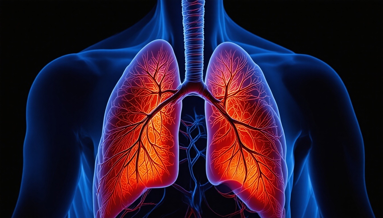
The human respiratory system is a marvel of biological engineering, responsible for the essential exchange of gases that sustains life. This system comprises a series of organs and structures that work in concert to bring oxygen into the body and expel carbon dioxide. Understanding the anatomy of the human respiratory system is crucial for students of medicine, biology, and allied health fields. In this comprehensive guide, we will delve into the structure of the human respiratory system, exploring the parts and functions of the structure of the human respiratory system, with a particular focus on the trachea, bronchioles, and alveoli. By the end, you will have a deep, academic-level understanding of how each component contributes to respiration.
Structure of the Human Respiratory System
The structure of the human respiratory system is anatomically divided into the upper respiratory tract and the lower respiratory tract. The upper tract includes the nose, nasal cavity, sinuses, and pharynx, while the lower tract comprises the larynx, trachea, bronchi, bronchioles, and alveoli. This hierarchical arrangement ensures that air is filtered, warmed, and humidified before reaching the delicate gas exchange surfaces.
From a functional perspective, the respiratory system can be categorized into conducting zones and respiratory zones. The conducting zone includes all structures that transport air to the respiratory zone, where gas exchange occurs. The respiratory zone consists of respiratory bronchioles, alveolar ducts, and alveoli. This division is critical for understanding how air moves and how oxygen and carbon dioxide are exchanged.
Embryologically, the respiratory system develops from the foregut endoderm, with the laryngotracheal groove forming the trachea and lung buds. This developmental origin explains the continuity between the digestive and respiratory tracts at the pharynx. Knowledge of this embryology aids in understanding congenital anomalies such as tracheoesophageal fistula.
Parts and Functions of the Structure of the Human Respiratory System
The parts and functions of the structure of the human respiratory system are intricately linked. Each component has a specialized role that contributes to the overall process of respiration. Below, we examine these parts in detail, from the nasal cavity to the alveoli.
Nasal Cavity
The nasal cavity is the primary entry point for air. It is lined with ciliated pseudostratified columnar epithelium that secretes mucus, trapping dust and pathogens. The rich blood supply warms the air, while the turbinates create turbulent airflow to maximize contact with mucus. The nasal cavity also houses olfactory receptors for smell.
Pharynx
The pharynx is a muscular tube that serves both respiratory and digestive functions. It is divided into nasopharynx, oropharynx, and laryngopharynx. The nasopharynx contains the pharyngeal tonsils (adenoids) and the openings of the Eustachian tubes. The oropharynx and laryngopharynx are common pathways for air and food.
Larynx
The larynx, or voice box, connects the pharynx to the trachea. It is composed of cartilages (thyroid, cricoid, and arytenoid) and contains the vocal cords. The epiglottis prevents food from entering the trachea during swallowing. The larynx also produces sound for speech.
Trachea
The trachea, commonly known as the windpipe, is a tubular structure about 10-12 cm long and 2 cm in diameter, extending from the larynx to the carina (the bifurcation point into the left and right main bronchi). It is a key component of the conducting zone, ensuring that air reaches the lungs efficiently.
The tracheal wall consists of four layers: mucosa, submucosa, hyaline cartilage, and adventitia. The mucosa is lined with ciliated pseudostratified columnar epithelium and goblet cells that produce mucus. The submucosa contains seromucous glands. The C-shaped hyaline cartilage rings (16-20 in number) provide structural support and prevent collapse, while the posterior wall is composed of smooth muscle (trachealis muscle) that allows for flexibility during swallowing.
The trachea is located anterior to the esophagus and extends from the level of C6 to T4-T5. It bifurcates at the carina, which is a sensitive area that triggers coughing when irritated. The right main bronchus is wider, shorter, and more vertical than the left, making it more susceptible to aspiration of foreign objects.
Bronchioles
Bronchioles are the smaller branches of the bronchial tree, typically less than 1 mm in diameter. They lack cartilage and glands, distinguishing them from bronchi. The bronchioles are part of both the conducting and respiratory zones, with terminal bronchioles being the last part of the conducting zone and respiratory bronchioles marking the beginning of the respiratory zone.
The walls of bronchioles are composed of smooth muscle and elastic fibers, allowing for bronchodilation and bronchoconstriction under autonomic control. Sympathetic stimulation causes dilation, while parasympathetic stimulation causes constriction. This regulation is crucial for conditions like asthma, where excessive constriction impairs airflow.
Each terminal bronchiole divides into several respiratory bronchioles, which then lead into alveolar ducts and alveolar sacs. The transition from conducting to respiratory zone is gradual, with respiratory bronchioles having occasional alveoli budding from their walls. This design maximizes the surface area for gas exchange.
Alveoli
Alveoli are the tiny air sacs where gas exchange occurs. There are approximately 300 million alveoli in the human lungs, providing a total surface area of about 70-100 square meters. Each alveolus is surrounded by a dense network of pulmonary capillaries, creating a thin barrier for diffusion.
The alveolar wall consists of two types of epithelial cells: type I pneumocytes (squamous) and type II pneumocytes (cuboidal). Type I cells cover 95% of the surface area and are extremely thin (0.2 μm) to facilitate gas diffusion. Type II cells produce pulmonary surfactant, a phospholipid-protein complex that reduces surface tension, preventing alveolar collapse (atelectasis).
Alveolar macrophages (dust cells) patrol the alveolar surfaces, phagocytizing inhaled particles and pathogens. The interalveolar septa contain elastic fibers that allow alveoli to expand during inhalation and recoil during exhalation. The close apposition of alveolar and capillary basement membranes forms the respiratory membrane, which is only 0.5 μm thick, enabling efficient O2 and CO2 exchange.
Gas Exchange and Transport
Gas exchange occurs across the respiratory membrane via simple diffusion. Oxygen diffuses from the alveoli (partial pressure ~100 mmHg) into the blood (partial pressure ~40 mmHg), while carbon dioxide diffuses in the opposite direction. This process is driven by concentration gradients and is influenced by factors such as membrane thickness, surface area, and ventilation-perfusion matching.
Oxygen is transported in the blood primarily bound to hemoglobin in red blood cells (98.5%), with a small amount dissolved in plasma. Carbon dioxide is transported in three forms: dissolved (5-10%), bound to hemoglobin as carbaminohemoglobin (20-30%), and as bicarbonate ions (60-70%) formed in red blood cells via carbonic anhydrase.
The efficiency of gas exchange is measured by parameters such as alveolar ventilation, dead space, and diffusion capacity. Alveolar ventilation is the volume of air reaching the alveoli per minute, while dead space refers to areas where no gas exchange occurs (e.g., conducting airways). The diffusion capacity of the lung for carbon monoxide (DLCO) is a clinical test to assess gas transfer.
Mechanics of Breathing
Breathing involves two phases: inspiration (inhalation) and expiration (exhalation). Inspiration is an active process driven by contraction of the diaphragm and external intercostal muscles, which expand the thoracic cavity and decrease intrapleural pressure, drawing air into the lungs. Expiration is normally passive, relying on elastic recoil of the lungs and chest wall.
During forced breathing, accessory muscles such as the sternocleidomastoid, scalenes, and pectoralis minor assist in inspiration, while internal intercostals and abdominal muscles aid in active expiration. The intrapleural pressure is always negative relative to atmospheric pressure, maintaining lung expansion. Pneumothorax occurs when air enters the pleural space, causing lung collapse.
Lung volumes and capacities are measured by spirometry. Key volumes include tidal volume (TV), inspiratory reserve volume (IRV), expiratory reserve volume (ERV), and residual volume (RV). Capacities are sums of volumes: total lung capacity (TLC), vital capacity (VC), inspiratory capacity (IC), and functional residual capacity (FRC). These measurements are critical for diagnosing pulmonary diseases.
Control of Respiration
Respiration is controlled by the respiratory center in the medulla oblongata and pons of the brainstem. The medullary respiratory center includes the dorsal respiratory group (DRG) and ventral respiratory group (VRG). The DRG primarily controls inspiration, while the VRG is involved in forced breathing. The pontine centers (pneumotaxic and apneustic) modulate the rhythm.
Chemoreceptors monitor blood pH, CO2, and O2 levels. Central chemoreceptors in the medulla are sensitive to changes in CSF pH (indirectly reflecting CO2), while peripheral chemoreceptors (carotid and aortic bodies) detect low O2, high CO2, and low pH. The primary drive for breathing is CO2 levels; hypercapnia stimulates increased ventilation.
Other factors influencing respiration include the Hering-Breuer reflex (stretch receptors in lungs prevent overinflation), baroreceptors, and voluntary control from the cerebral cortex. During exercise, ventilation increases dramatically to meet metabolic demands, with mechanisms involving feed-forward signals from motor cortex and feedback from chemoreceptors and mechanoreceptors.
Clinical Correlations
Understanding the anatomy of the respiratory system is essential for diagnosing and treating respiratory disorders. Common conditions include asthma, chronic obstructive pulmonary disease (COPD), pneumonia, and lung cancer. Asthma involves bronchoconstriction and inflammation of bronchioles, while COPD encompasses emphysema (alveolar destruction) and chronic bronchitis.
Pneumonia is an infection of the alveoli, leading to fluid accumulation and impaired gas exchange. Lung cancer often arises from bronchial epithelium and can obstruct airways. Other conditions like pulmonary embolism (blockage of pulmonary artery) and acute respiratory distress syndrome (ARDS) affect gas exchange at the alveolar-capillary level.
Diagnostic tools include chest X-ray, CT scan, bronchoscopy, pulmonary function tests (PFTs), and arterial blood gas analysis. Treatment may involve bronchodilators, corticosteroids, oxygen therapy, mechanical ventilation, or surgery. Knowledge of respiratory anatomy guides interventions like endotracheal intubation (trachea) and thoracentesis (pleural space).
Detailed Anatomy of the Nasal Cavity
The nasal cavity is divided into right and left halves by the nasal septum, which is composed of the perpendicular plate of the ethmoid bone, vomer, and septal cartilage. Each lateral wall contains three bony projections called conchae (superior, middle, and inferior), which increase surface area and create turbulent airflow. The epithelium lining the nasal cavity is primarily respiratory epithelium, except in the olfactory region where specialized olfactory epithelium is present.
The nasal cavity receives blood supply from branches of the ophthalmic and maxillary arteries, including the sphenopalatine artery, which is a common site of epistaxis (nosebleed). Innervation comes from the olfactory nerve (CN I) for smell and branches of the trigeminal nerve (CN V) for general sensation. The nasolacrimal duct drains tears from the eye into the inferior meatus.
Functions of the nasal cavity include filtration, warming, and humidification of inhaled air. The mucus layer traps particles, and cilia move the mucus toward the pharynx for swallowing. The rich vascular network warms the air to body temperature, and the turbinates increase contact time. Additionally, the nasal cavity contributes to resonance during speech.
Detailed Anatomy of the Pharynx
The pharynx extends from the base of the skull to the level of C6, where it continues as the esophagus. It is divided into three regions: nasopharynx (posterior to the nasal cavity), oropharynx (posterior to the oral cavity), and laryngopharynx (posterior to the larynx). The nasopharynx is lined by respiratory epithelium, while the oropharynx and laryngopharynx are lined by stratified squamous epithelium due to abrasion from food.
The pharyngeal walls consist of four layers: mucosa, submucosa (pharyngobasilar fascia), muscular layer (constrictor muscles), and buccopharyngeal fascia. The superior, middle, and inferior constrictor muscles overlap to propel food toward the esophagus. The pharyngeal plexus (CN IX and X) provides motor innervation, while sensory innervation comes from CN IX and X.
Lymphoid tissue in the pharynx forms Waldeyer’s ring, which includes the pharyngeal tonsil (adenoids), tubal tonsils, palatine tonsils, and lingual tonsil. These tonsils are part of the immune system, trapping pathogens. The pharynx also plays a role in equalizing pressure in the middle ear via the Eustachian tube openings in the nasopharynx.
Detailed Anatomy of the Larynx
The larynx is located at the level of C4-C6 and is composed of nine cartilages: three paired (arytenoid, corniculate, cuneiform) and three unpaired (thyroid, cricoid, epiglottis). The thyroid cartilage is the largest and forms the Adam’s apple. The cricoid cartilage is a complete ring, and the arytenoid cartilages articulate with the cricoid and attach to the vocal cords.
The intrinsic muscles of the larynx control tension and position of the vocal cords. The cricothyroid muscle tenses the cords, while the thyroarytenoid muscle (vocalis) relaxes them. The posterior cricoarytenoid muscles abduct the cords, and the lateral cricoarytenoid and interarytenoid muscles adduct them. The recurrent laryngeal nerve (branch of CN X) innervates all intrinsic muscles except the cricothyroid (innervated by the external branch of the superior laryngeal nerve).
The laryngeal cavity is divided into three regions: vestibule (above the vestibular folds), ventricle (between vestibular and vocal folds), and infraglottic cavity (below the vocal folds). The glottis is the opening between the vocal cords. During phonation, the vocal cords adduct and vibrate, producing sound. The pitch is determined by tension and length of the cords.
Detailed Anatomy of the Trachea
The trachea is a fibromuscular tube supported by 16-20 C-shaped hyaline cartilage rings. The open ends of the rings are posterior, where the trachealis muscle (smooth muscle) connects them. This arrangement allows the esophagus to expand during swallowing. The trachea is lined by ciliated pseudostratified columnar epithelium with goblet cells, which produce mucus that traps particles, and cilia that beat upward to propel mucus toward the pharynx.
The blood supply to the trachea comes from the inferior thyroid artery (upper part) and bronchial arteries (lower part). Venous drainage is to the inferior thyroid and brachiocephalic veins. Lymphatic drainage goes to pretracheal and paratracheal lymph nodes. The trachea is innervated by the recurrent laryngeal nerves and sympathetic fibers from the cervical ganglia.
Clinical significance: Tracheostomy is a surgical procedure to create an opening in the trachea to bypass an obstruction. The trachea can be compressed by goiter or tumors. Tracheal stenosis may occur after prolonged intubation. The carina is a sensitive area; irritation here triggers a strong cough reflex.
Detailed Anatomy of the Bronchi and Bronchioles
The trachea bifurcates at the carina into the right and left main bronchi. The right main bronchus is wider, shorter (2.5 cm), and more vertical, making it the most common site for aspiration of foreign bodies. The left main bronchus is narrower, longer (5 cm), and more horizontal, passing under the aortic arch. Each main bronchus enters the hilum of the respective lung.
Within the lungs, the main bronchi divide into lobar bronchi (three on the right, two on the left), then segmental bronchi, and further into bronchioles. The bronchial tree undergoes about 23 generations of branching. As branching progresses, cartilage decreases and smooth muscle increases. Bronchioles are less than 1 mm in diameter and lack cartilage and mucous glands.
The terminal bronchioles are the last part of the conducting zone. Each terminal bronchiole divides into respiratory bronchioles, which have alveoli budding from their walls. The respiratory bronchioles lead to alveolar ducts and alveolar sacs. The walls of bronchioles contain Clara cells, which secrete surfactant-like substances and detoxify inhaled toxins. The smooth muscle of bronchioles is under autonomic control; bronchoconstriction in asthma is a major clinical concern.
Detailed Anatomy of the Alveoli
Alveoli are polyhedral sacs with a diameter of about 200-300 μm. They are clustered in groups called alveolar sacs, which open from alveolar ducts. The interalveolar septa contain capillaries, elastic fibers, collagen, and fibroblasts. Pores of Kohn are small openings between adjacent alveoli that allow collateral ventilation and equalization of pressure.
Type I pneumocytes are extremely thin squamous cells that cover 95% of the alveolar surface. They are joined by tight junctions and have few organelles. Type II pneumocytes are cuboidal cells that produce surfactant, a mixture of phospholipids (mainly dipalmitoylphosphatidylcholine) and proteins (SP-A, SP-B, SP-C, SP-D). Surfactant reduces surface tension, preventing alveolar collapse and reducing the work of breathing.
Alveolar macrophages are derived from blood monocytes and migrate into the alveoli. They phagocytize inhaled particles, bacteria, and debris. They can be seen in sputum samples as “dust cells”. The respiratory membrane consists of the alveolar epithelium, basement membrane, capillary basement membrane, and capillary endothelium. The total thickness is only 0.5 μm, allowing rapid diffusion.
Pleura and Pleural Cavity
The pleura is a serous membrane that surrounds the lungs. The visceral pleura adheres to the lung surface, while the parietal pleura lines the thoracic cavity. The two layers are continuous at the hilum and form the pleural cavity, which contains a thin film of serous fluid (about 10-20 mL) that reduces friction during breathing.
The parietal pleura is divided into four parts: costal (ribs), diaphragmatic (diaphragm), mediastinal (mediastinum), and cervical (cupula, extending above the first rib). The pleural cavity has recesses where the lungs don’t fully occupy, such as the costodiaphragmatic recess (between diaphragm and ribs) and costomediastinal recess. These recesses allow lung expansion during inspiration.
Innervation of the parietal pleura is by intercostal nerves (costal part) and phrenic nerve (diaphragmatic and mediastinal parts), which explains referred pain to the shoulder in diaphragmatic irritation. The visceral pleura is innervated by autonomic fibers and is insensitive to pain. Pleurisy (inflammation of the pleura) causes sharp pain on inspiration.
Lungs: Lobes, Fissures, and Hilum
The right lung has three lobes: superior, middle, and inferior, separated by the horizontal and oblique fissures. The left lung has two lobes: superior and inferior, separated by the oblique fissure. The left lung also has a cardiac notch to accommodate the heart. Each lobe is divided into bronchopulmonary segments, which are functionally independent units supplied by a segmental bronchus and artery.
The hilum is the medial region where the bronchus, pulmonary artery, pulmonary veins, bronchial vessels, nerves, and lymphatics enter and exit the lung. The arrangement at the hilum is: pulmonary artery superior, bronchus posterior, and pulmonary veins anterior (remember “A-B-V”). The root of the lung is the structures connecting the lung to the mediastinum.
The bronchopulmonary segments are 10 in the right lung and 8-10 in the left lung. Each segment is separated by connective tissue septa and can be surgically removed without affecting adjacent segments. Knowledge of segmental anatomy is crucial for locating lesions and planning resections.
Blood Supply and Lymphatics of the Lungs
The lungs have a dual blood supply: the pulmonary circulation for gas exchange and the bronchial circulation for nourishment of lung tissue. The pulmonary arteries carry deoxygenated blood from the right ventricle to the lungs, branching along the bronchial tree. The pulmonary veins carry oxygenated blood back to the left atrium. The bronchial arteries (from the thoracic aorta) supply the bronchi, pleura, and supporting tissues.
Lymphatic drainage of the lungs begins with lymph capillaries in the interlobular septa and around bronchioles. Lymph flows to hilar lymph nodes, then to tracheobronchial, paratracheal, and mediastinal nodes. The right lymphatic duct and thoracic duct drain lymph into the venous system. Lymphatic spread is a common route for lung cancer metastasis.
Nerve supply to the lungs comes from the pulmonary plexus, which contains sympathetic (T1-T5) and parasympathetic (vagus) fibers. Sympathetic stimulation causes bronchodilation and vasoconstriction, while parasympathetic stimulation causes bronchoconstriction and vasodilation. Afferent fibers carry sensory information from stretch receptors and chemoreceptors.
Developmental Anatomy and Congenital Anomalies
The respiratory system develops from the foregut at around 4 weeks of gestation. The laryngotracheal groove appears on the ventral side of the foregut, which deepens to form the laryngotracheal tube. The distal end of the tube forms the lung buds, which branch into the bronchial tree. The surrounding mesenchyme gives rise to cartilage, smooth muscle, and connective tissue.
Congenital anomalies include tracheoesophageal fistula (abnormal connection between trachea and esophagus), laryngomalacia (soft laryngeal cartilage), and congenital diaphragmatic hernia (abdominal organs herniate into chest, compressing lungs). Pulmonary hypoplasia is underdevelopment of lung tissue, often associated with oligohydramnios. Cystic adenomatoid malformation is a hamartomatous lesion of the lung.
Understanding embryology helps explain the segmental anatomy and variations in bronchial branching. for instance, the right upper lobe bronchus often arises directly from the trachea (tracheal bronchus) in some individuals. Such variations are important for bronchoscopy and surgical planning.
Age-Related Changes in the Respiratory System
With aging, the respiratory system undergoes several changes. The chest wall becomes stiffer due to calcification of costal cartilages and decreased elasticity. The diaphragm weakens, reducing maximal inspiratory and expiratory pressures. Lung elasticity decreases due to loss of elastin fibers, leading to increased compliance and air trapping (senile emphysema).
Alveolar surface area decreases by about 20% by age 70, reducing gas exchange efficiency. The number of alveoli remains relatively constant, but the size increases. The immune function declines, increasing susceptibility to infections. The mucociliary clearance becomes less effective, and the cough reflex weakens.
Pulmonary function tests show a decline in FEV1 (forced expiratory volume in 1 second) and FVC (forced vital capacity) with age. The residual volume increases, and the diffusion capacity decreases. These changes contribute to the increased prevalence of respiratory diseases in the elderly, such as pneumonia and COPD.
Respiratory System in Exercise
During exercise, the respiratory system must meet increased oxygen demand and eliminate excess carbon dioxide. Ventilation can increase from about 6 L/min at rest to over 100 L/min in elite athletes. This is achieved by increasing tidal volume and respiratory rate. The diaphragm and accessory muscles work harder.
Gas exchange efficiency improves due to better ventilation-perfusion matching and increased pulmonary capillary blood flow. Oxygen diffusion capacity increases as more capillaries are recruited. but, at very high intensities, the respiratory muscles may fatigue, limiting performance. Respiratory muscle training can improve exercise tolerance.
Exercise-induced bronchoconstriction (EIB) occurs in some individuals, especially in cold, dry air. It is thought to be due to airway drying and cooling, leading to mast cell degranulation. Treatment includes inhaled bronchodilators before exercise.
Respiratory System and Altitude
At high altitude, the partial pressure of oxygen in the air decreases, leading to hypobaric hypoxia. The body adapts through acclimatization, which includes increased ventilation (hyperventilation), increased heart rate, and increased red blood cell production (erythropoiesis) over days to weeks. The hypoxic ventilatory response is mediated by peripheral chemoreceptors.
Acute mountain sickness (AMS) can occur at altitudes above 2,500 m, with symptoms of headache, nausea, and fatigue. Severe forms include high-altitude pulmonary edema (HAPE) and high-altitude cerebral edema (HACE). HAPE is characterized by pulmonary hypertension and fluid leakage into alveoli, while HACE involves brain swelling.
Long-term adaptation in high-altitude populations includes larger lung volumes, increased capillary density, and genetic changes in oxygen sensing pathways. Understanding these adaptations provides insights into respiratory physiology and disease.
Conclusion
In conclusion, the anatomy of the human respiratory system is a complex and highly organized network of structures that facilitate the vital process of gas exchange. From the nasal cavity to the alveoli, each component plays a specific role in conducting air, filtering impurities, and enabling diffusion. The trachea provides a rigid airway, bronchioles regulate airflow, and alveoli offer an immense surface area for gas exchange. Understanding these parts and functions is fundamental for any student of human biology. This guide has provided a detailed exploration, integrating structure with function and clinical relevance. Mastery of this material will serve as a solid foundation for further study in physiology, pathology, and clinical medicine.
Frequently Asked Questions
What is the main function of the trachea?
The trachea serves as the main conduit for air from the larynx to the bronchi. It provides a rigid yet flexible airway, thanks to its C-shaped cartilage rings, and its ciliated epithelium helps trap and expel debris.
How do bronchioles differ from bronchi?
Bronchioles are smaller than bronchi (less than 1 mm diameter) and lack cartilage and glands. They have more smooth muscle, allowing for significant changes in diameter. Bronchioles are part of both conducting and respiratory zones.
What is the role of alveoli in gas exchange?
Alveoli are the microscopic air sacs where oxygen and carbon dioxide are exchanged between air and blood. Their thin walls (0.2 μm) and large surface area (70-100 m²) make diffusion highly efficient.
What causes the lungs to stick to the chest wall?
The pleural cavity contains a thin layer of fluid that creates surface tension, adhering the visceral pleura (lung) to the parietal pleura (chest wall). This negative intrapleural pressure keeps the lungs expanded.
How is breathing controlled?
Breathing is controlled by the respiratory center in the brainstem, which responds to chemoreceptor input (CO2, O2, pH). The medulla generates the basic rhythm, while the pons modulates it.
“,
“excerpt”: “Detailed academic notes on the anatomy of the human respiratory system, covering the trachea, bronchioles, alveoli, and all parts with functions, gas exchange, and clinical correlations.”
}


Leave a Reply