
Introduction
Aerobic respiration is the cornerstone of cellular energy metabolism in eukaryotic organisms. It is a highly efficient biochemical pathway that converts the chemical energy stored in nutrients, primarily glucose, into adenosine triphosphate (ATP), the universal energy currency of the cell. The overall process is exergonic, releasing energy that is harnessed to drive endergonic cellular processes. Understanding the biochemistry of aerobic respiration is crucial for fields ranging from medicine to biotechnology. This comprehensive guide delves into the intricate molecular mechanisms, the subcellular sites, and the regulatory aspects of aerobic respiration, with a focus on the mitochondria as the primary site. We will explore the four main stages: glycolysis, the link reaction, the Krebs cycle, and oxidative phosphorylation, each with its unique set of enzymes, intermediates, and energy yields. By the end of this article, you will have a deep appreciation for how cells extract maximum ATP from glucose, the role of oxygen as the final electron acceptor, and the elegant coupling of electron transport to ATP synthesis. This knowledge forms the foundation for understanding metabolic disorders, exercise physiology, and the evolution of life on an oxygen-rich planet.
The Process and Site of Aerobic Respiration
Aerobic respiration is a metabolic process that requires molecular oxygen (O₂) to produce ATP from organic fuels. The overall process can be summarized as the complete oxidation of glucose to carbon dioxide and water, with the concomitant reduction of oxygen to water. This process occurs in distinct cellular compartments, each specialized for specific biochemical reactions. The initial stage, glycolysis, takes place in the cytosol, where glucose is partially oxidized to pyruvate. but, the majority of ATP production occurs within the mitochondria, which are often referred to as the powerhouses of the cell. The mitochondria are double-membrane-bound organelles with a highly folded inner membrane called cristae, which increases the surface area for electron transport chain complexes and ATP synthase. The matrix, the fluid-filled space inside the inner membrane, houses the enzymes of the Krebs cycle and the link reaction. Thus, the site of aerobic respiration is predominantly the mitochondria, but it begins in the cytosol. The process is tightly regulated to meet the energy demands of the cell, and its efficiency is reflected in the net yield of approximately 30-32 ATP molecules per glucose molecule under ideal conditions. This high yield is due to the complete oxidation of glucose and the proton gradient-driven ATP synthesis in oxidative phosphorylation.
Word and Symbol Equations for Aerobic Respiration
The overall chemical equation for aerobic respiration succinctly captures the transformation of glucose and oxygen into carbon dioxide, water, and energy. The word equation is: Glucose + Oxygen → Carbon Dioxide + Water + Energy (as ATP and heat). The balanced symbol equation is: C₆H₁₂O₆ + 6O₂ → 6CO₂ + 6H₂O + Energy (approximately 30-32 ATP). This equation represents the complete combustion of glucose, but in reality, the process occurs through a series of enzyme-catalyzed steps. The energy released isn’t all captured as ATP; some is dissipated as heat, which helps maintain body temperature in endotherms. The symbol equation indicates the stoichiometry: one molecule of glucose requires six molecules of oxygen to produce six molecules of carbon dioxide and six molecules of water. but, this net equation doesn’t reveal the intermediate steps, such as the production of NADH and FADH₂, which carry electrons to the electron transport chain. The actual ATP yield isn’t a fixed number because it depends on the efficiency of the proton motive force and the leakage of protons across the inner mitochondrial membrane. Nevertheless, the equation provides a valuable summary of the overall redox reaction where glucose is oxidized and oxygen is reduced.
Mitochondria as the Site in Cells
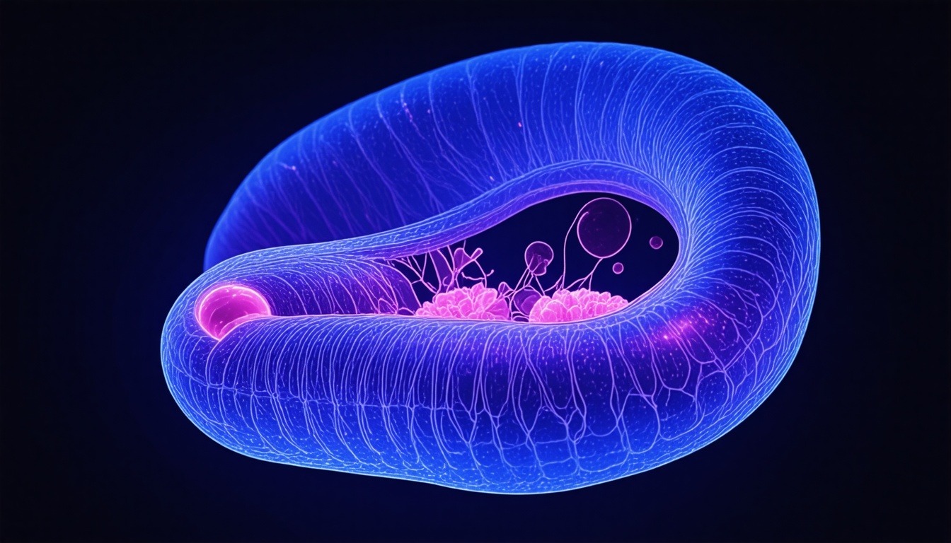
Mitochondria are double-membrane-bound organelles found in the cytoplasm of eukaryotic cells. Their structure is exquisitely adapted for aerobic respiration. The outer mitochondrial membrane is permeable to small molecules due to the presence of porins, while the inner membrane is highly impermeable and contains specific transport proteins. The inner membrane is folded into cristae, which dramatically increase its surface area, housing the electron transport chain complexes (I-IV) and ATP synthase. The intermembrane space, located between the outer and inner membranes, is where protons accumulate to create a proton gradient. The mitochondrial matrix contains the enzymes for the Krebs cycle, the link reaction, and fatty acid oxidation, as well as mitochondrial DNA and ribosomes. This compartmentalization allows for the efficient coupling of electron transport to ATP synthesis via chemiosmosis. The number of mitochondria per cell varies depending on the cell’s energy demand; for instance, muscle cells and neurons have high mitochondrial densities. Mitochondria are also dynamic organelles, undergoing fusion and fission to maintain their function and quality control. Dysfunctions in mitochondria are linked to numerous diseases, including neurodegenerative disorders, metabolic syndromes, and aging. Thus, understanding mitochondria as the site of aerobic respiration is fundamental to cellular biology and medicine.
Glycolysis: The First Stage
Glycolysis is the initial metabolic pathway of aerobic respiration, occurring in the cytosol. It involves the breakdown of one molecule of glucose (6 carbons) into two molecules of pyruvate (3 carbons each), with a net production of 2 ATP and 2 NADH. The pathway consists of ten enzyme-catalyzed steps, divided into two phases: the energy investment phase (steps 1-5) and the energy payoff phase (steps 6-10). In the investment phase, two ATP molecules are consumed to phosphorylate glucose, forming fructose-1,6-bisphosphate, which is then split into two triose phosphates: glyceraldehyde-3-phosphate (G3P) and dihydroxyacetone phosphate (DHAP). Only G3P continues in glycolysis; DHAP is isomerized to G3P. In the payoff phase, each G3P is oxidized and phosphorylated to produce 1,3-bisphosphoglycerate, which donates a phosphate to ADP to form ATP via substrate-level phosphorylation. Subsequent steps lead to the production of pyruvate. The net reaction is: Glucose + 2 NAD⁺ + 2 ADP + 2 Pi → 2 Pyruvate + 2 NADH + 2 H⁺ + 2 ATP + 2 H₂O. Glycolysis doesn’t require oxygen, so it can occur under anaerobic conditions, but for aerobic respiration to continue, the pyruvate must enter the mitochondria and be further oxidized.
Key Enzymes of Glycolysis
The key regulatory enzymes in glycolysis are hexokinase, phosphofructokinase-1 (PFK-1), and pyruvate kinase. Hexokinase catalyzes the first step: phosphorylation of glucose to glucose-6-phosphate, trapping glucose inside the cell. PFK-1 is the most important control point; it catalyzes the irreversible phosphorylation of fructose-6-phosphate to fructose-1,6-bisphosphate. PFK-1 is allosterically activated by AMP and fructose-2,6-bisphosphate, and inhibited by ATP and citrate. Pyruvate kinase catalyzes the final step, transferring a phosphate from phosphoenolpyruvate to ADP, yielding pyruvate and ATP. It is activated by fructose-1,6-bisphosphate and inhibited by ATP and alanine. These regulatory mechanisms ensure that glycolysis is upregulated when energy demand is high and downregulated when ATP is abundant.
Energy Yield of Glycolysis
Glycolysis produces a net gain of 2 ATP and 2 NADH per glucose molecule. Under aerobic conditions, the NADH can be shuttled into the mitochondria and oxidized via the electron transport chain, yielding additional ATP (about 1.5-2.5 ATP per NADH, depending on the shuttle system). but, the ATP produced directly in glycolysis is via substrate-level phosphorylation, which is independent of oxygen. This makes glycolysis a rapid source of ATP, especially in cells lacking mitochondria (e.g., red blood cells) or under temporary oxygen deficits (e.g., during intense exercise). The pyruvate produced is transported into the mitochondrial matrix for further oxidation in the link reaction.
The Link Reaction (Pyruvate Decarboxylation)
Before pyruvate can enter the Krebs cycle, it must be converted into acetyl-CoA in a process called the link reaction, which occurs in the mitochondrial matrix. This irreversible reaction is catalyzed by the pyruvate dehydrogenase complex, a multi-enzyme complex that requires five cofactors: thiamine pyrophosphate (TPP), lipoic acid, coenzyme A (CoA), FAD, and NAD⁺. The overall reaction is: Pyruvate + CoA + NAD⁺ → Acetyl-CoA + CO₂ + NADH + H⁺. This reaction involves three steps: decarboxylation (removal of CO₂), oxidation (transfer of electrons to NAD⁺ to form NADH), and transfer of the acetyl group to CoA. The pyruvate dehydrogenase complex is tightly regulated by covalent modification (phosphorylation inactivates it) and allosteric effectors (e.g., ATP, NADH, and acetyl-CoA inhibit it; ADP and Ca²⁺ activate it). This regulation ensures that the link reaction only proceeds when the cell needs energy and when the Krebs cycle is ready to accept acetyl-CoA. The CO₂ released is a waste product that is exhaled. The NADH produced is a high-energy electron carrier that will donate electrons to the electron transport chain, contributing to ATP synthesis.
The Krebs Cycle (Citric Acid Cycle)
The Krebs cycle, also known as the citric acid cycle or tricarboxylic acid cycle, takes place in the mitochondrial matrix. It is a central metabolic hub that oxidizes acetyl-CoA completely to CO₂, generating high-energy electron carriers (NADH and FADH₂) and a small amount of ATP (or GTP). The cycle begins with the condensation of acetyl-CoA (2 carbons) with oxaloacetate (4 carbons) to form citrate (6 carbons), catalyzed by citrate synthase. Citrate is then isomerized to isocitrate, which undergoes oxidative decarboxylation to α-ketoglutarate (5 carbons), producing NADH and CO₂. α-Ketoglutarate is further decarboxylated to succinyl-CoA (4 carbons), generating another NADH and CO₂. Succinyl-CoA is converted to succinate, producing GTP (which can be converted to ATP) via substrate-level phosphorylation by succinyl-CoA synthetase. Succinate is then oxidized to fumarate, producing FADH₂ by succinate dehydrogenase (which is actually part of the electron transport chain). Fumarate is hydrated to malate, and malate is oxidized to oxaloacetate, producing another NADH. The net reaction per acetyl-CoA is: Acetyl-CoA + 3 NAD⁺ + FAD + GDP + Pi → 2 CO₂ + 3 NADH + FADH₂ + GTP + 2 H⁺ + CoA. Since each glucose yields two pyruvates and thus two acetyl-CoA, the Krebs cycle turns twice per glucose, producing 6 NADH, 2 FADH₂, and 2 GTP (equivalent to 2 ATP). The cycle is regulated at key irreversible steps: citrate synthase, isocitrate dehydrogenase, and α-ketoglutarate dehydrogenase, which are inhibited by high levels of ATP, NADH, and succinyl-CoA, and activated by ADP and Ca²⁺.
Oxidative Phosphorylation: Electron Transport Chain and Chemiosmosis
Oxidative phosphorylation is the final and most productive stage of aerobic respiration, occurring in the inner mitochondrial membrane. It involves two coupled processes: the electron transport chain (ETC) and chemiosmosis. The ETC consists of four protein complexes (I, II, III, IV) and two mobile carriers (ubiquinone and cytochrome c). Electrons from NADH and FADH₂ are passed through these complexes, releasing energy that is used to pump protons (H⁺) from the matrix to the intermembrane space, creating an electrochemical gradient (proton motive force). Complex I (NADH dehydrogenase) accepts electrons from NADH and transfers them to ubiquinone, pumping 4 H⁺. Complex II (succinate dehydrogenase) accepts electrons from FADH₂ (from the Krebs cycle) and transfers them to ubiquinone, but doesn’t pump protons. Complex III (cytochrome bc1 complex) transfers electrons from ubiquinol to cytochrome c, pumping 4 H⁺. Complex IV (cytochrome c oxidase) transfers electrons from cytochrome c to oxygen, the final electron acceptor, forming water and pumping 2 H⁺. The overall reaction for the ETC is: NADH + 11 H⁺ (matrix) + ½ O₂ → NAD⁺ + 10 H⁺ (intermembrane) + H₂O (for NADH); for FADH₂: FADH₂ + 6 H⁺ (matrix) + ½ O₂ → FAD + 6 H⁺ (intermembrane) + H₂O. The proton gradient then drives ATP synthesis as protons flow back into the matrix through ATP synthase (Complex V), a molecular turbine that couples proton flow to the phosphorylation of ADP. This process is called chemiosmosis. Typically, 3-4 H⁺ are required to produce one ATP. The theoretical ATP yield from oxidative phosphorylation is about 2.5 ATP per NADH and 1.5 ATP per FADH₂, but actual yields are lower due to proton leakage and other inefficiencies. The chemiosmotic theory was proposed by Peter Mitchell and is a cornerstone of bioenergetics.
Complex I: NADH Dehydrogenase
Complex I is a large L-shaped enzyme composed of 45 subunits in mammals. It catalyzes the oxidation of NADH to NAD⁺, transferring two electrons to ubiquinone (Q) to form ubiquinol (QH₂). The reaction is: NADH + Q + 5 H⁺ (matrix) → NAD⁺ + QH₂ + 4 H⁺ (intermembrane). The energy released from electron transfer drives the pumping of four protons across the membrane. Complex I is a major site of reactive oxygen species (ROS) production when it is inefficient or inhibited. Mutations in complex I subunits are associated with mitochondrial diseases such as Leigh syndrome.
Complex II: Succinate Dehydrogenase
Complex II is the only ETC complex that is also part of the Krebs cycle. It catalyzes the oxidation of succinate to fumarate, reducing FAD to FADH₂, which then transfers electrons to ubiquinone. No protons are pumped at this complex, so it contributes less to the proton gradient. Complex II is a small complex with four subunits and contains iron-sulfur clusters. It isn’t a major source of ROS but can be involved in certain metabolic disorders.
Complex III: Cytochrome bc1 Complex
Complex III catalyzes the transfer of electrons from ubiquinol to cytochrome c, coupled to proton pumping. It operates via the Q cycle, which enhances proton translocation. For each pair of electrons, two ubiquinol molecules are oxidized, and one ubiquinone is regenerated, with the net pumping of 4 H⁺. The reaction is: QH₂ + 2 cytochrome c (oxidized) + 2 H⁺ (matrix) → Q + 2 cytochrome c (reduced) + 4 H⁺ (intermembrane). Complex III is a dimer and contains cytochrome b, cytochrome c1, and an iron-sulfur protein. It is a target of many inhibitors, such as antimycin A.
Complex IV: Cytochrome c Oxidase
Complex IV is the terminal oxidase, transferring electrons from cytochrome c to oxygen, reducing it to water. It pumps 2 H⁺ per pair of electrons. The reaction is: 4 cytochrome c (reduced) + 8 H⁺ (matrix) + O₂ → 4 cytochrome c (oxidized) + 4 H⁺ (intermembrane) + 2 H₂O. Complex IV contains copper and heme centers (cytochrome a and a3). It is inhibited by cyanide and carbon monoxide, which bind to the heme a3-CuB center, blocking oxygen reduction and causing rapid cell death.
ATP Synthase (Complex V)
ATP synthase is a molecular motor that uses the proton gradient to synthesize ATP. It consists of two main parts: the F₁ head (catalytic) and the F₀ base (proton channel). As protons flow through F₀, they cause the rotation of a central stalk (γ subunit), which induces conformational changes in the F₁ head that allow ADP and Pi to bind and form ATP. The rotation is driven by the proton motive force, and it is estimated that 3-4 protons are needed to produce one ATP. The overall reaction is: ADP + Pi + n H⁺ (intermembrane) → ATP + H₂O + n H⁺ (matrix). ATP synthase can also work in reverse to pump protons, a process used by some bacteria to maintain their proton gradient. It is a highly conserved enzyme found in mitochondria, chloroplasts, and bacteria.
Regulation of Aerobic Respiration
Aerobic respiration is tightly regulated to match the cell’s energy demands. The key control points are the allosteric enzymes in glycolysis (PFK-1), the link reaction (pyruvate dehydrogenase complex), and the Krebs cycle (isocitrate dehydrogenase and α-ketoglutarate dehydrogenase). These enzymes are inhibited by high levels of ATP, NADH, and citrate, and activated by ADP, AMP, and NAD⁺. Additionally, the availability of oxygen is a critical factor; under hypoxic conditions, cells shift to anaerobic glycolysis, producing lactate. Hormonal regulation also occurs; for instance, insulin promotes glucose uptake and glycolysis, while glucagon and epinephrine stimulate glycogen breakdown and gluconeogenesis. Calcium ions (Ca²⁺) activate several dehydrogenases in the Krebs cycle and the pyruvate dehydrogenase complex, linking energy production to muscle contraction and neuronal activity. The regulation ensures that ATP production is finely tuned to cellular needs, preventing wasteful overproduction and maintaining redox balance.
Energy Yield and Efficiency
The theoretical maximum ATP yield from one molecule of glucose during aerobic respiration is about 30-32 ATP. This includes: 2 ATP from glycolysis (substrate-level), 2 ATP from the Krebs cycle (as GTP), and about 26-28 ATP from oxidative phosphorylation (from 10 NADH and 2 FADH₂). The actual yield is lower due to proton leakage, the cost of transporting NADH from glycolysis into the mitochondria (which consumes ATP equivalents), and the inefficiency of the electron transport chain. The overall efficiency of aerobic respiration is about 40%, meaning that 40% of the energy released from glucose oxidation is captured as ATP, while the rest is dissipated as heat. This efficiency is higher than that of anaerobic respiration (about 2% from glycolysis alone) and is why aerobic organisms can sustain complex, energy-intensive activities. The high ATP yield is essential for processes such as muscle contraction, active transport, and biosynthesis.
Comparison with Anaerobic Respiration
Aerobic respiration is far more efficient than anaerobic respiration, which doesn’t use oxygen and only partially oxidizes glucose. Anaerobic respiration in humans produces lactate and only 2 ATP per glucose via glycolysis alone. In yeast, anaerobic respiration produces ethanol and CO₂, also yielding 2 ATP. The absence of oxygen means that the NADH produced in glycolysis can’t be reoxidized by the electron transport chain; instead, it is recycled by reducing pyruvate to lactate or ethanol. This allows glycolysis to continue but wastes the energy stored in NADH. Aerobic respiration, by contrast, fully oxidizes glucose to CO₂ and H₂O, extracting the maximum possible energy. The ability to use oxygen as the final electron acceptor allows for a much larger proton gradient and thus more ATP. The evolution of aerobic respiration was a major milestone, enabling the development of complex multicellular life.
Clinical Relevance and Disorders
Defects in aerobic respiration can lead to a variety of mitochondrial diseases, which often affect tissues with high energy demands, such as muscle, brain, and heart. These disorders can be caused by mutations in mitochondrial DNA (mtDNA) or nuclear genes encoding ETC subunits or assembly factors. Common symptoms include muscle weakness, exercise intolerance, neurological deficits, and lactic acidosis. Examples include Leigh syndrome, MELAS (Mitochondrial Encephalopathy, Lactic Acidosis, and Stroke-like episodes), and Kearns-Sayre syndrome. Additionally, impaired aerobic respiration is implicated in aging, neurodegenerative diseases (e.g., Parkinson’s, Alzheimer’s), and cancer (the Warburg effect, where cancer cells rely on glycolysis even in the presence of oxygen). Understanding the biochemistry of aerobic respiration is crucial for developing therapeutic strategies, such as antioxidants to reduce ROS damage, or metabolic modulators to bypass defective ETC complexes.
Frequently Asked Questions
What is the main purpose of aerobic respiration?
The main purpose of aerobic respiration is to produce ATP, the primary energy currency of the cell, by completely oxidizing glucose in the presence of oxygen. This process provides the energy needed for cellular work, including biosynthesis, active transport, and mechanical work.
Where does aerobic respiration occur?
Aerobic respiration begins in the cytosol with glycolysis, but the majority of ATP production occurs in the mitochondria. The link reaction and Krebs cycle take place in the mitochondrial matrix, while oxidative phosphorylation occurs in the inner mitochondrial membrane.
What is the net ATP yield from aerobic respiration?
The net ATP yield from one molecule of glucose is approximately 30-32 ATP molecules under ideal conditions. This includes 2 ATP from glycolysis, 2 ATP (as GTP) from the Krebs cycle, and about 26-28 ATP from oxidative phosphorylation.
Why is oxygen necessary for aerobic respiration?
Oxygen serves as the final electron acceptor in the electron transport chain. It accepts electrons and protons to form water, allowing the chain to continue functioning. Without oxygen, the electron transport chain would halt, and oxidative phosphorylation would cease, drastically reducing ATP production.
What are the main stages of aerobic respiration?
The main stages are glycolysis (cytosol), the link reaction (mitochondrial matrix), the Krebs cycle (mitochondrial matrix), and oxidative phosphorylation (inner mitochondrial membrane). Each stage involves specific enzymes and produces intermediates that feed into the next stage.
How is aerobic respiration regulated?
Aerobic respiration is regulated primarily by the energy status of the cell. Key enzymes like phosphofructokinase-1, pyruvate dehydrogenase, and isocitrate dehydrogenase are inhibited by high ATP and NADH and activated by ADP and AMP. Hormones and calcium ions also modulate the pathway.
What happens if aerobic respiration is impaired?
Impairment of aerobic respiration can lead to mitochondrial diseases, characterized by muscle weakness, neurological problems, and lactic acidosis. It is also associated with aging and certain cancers. Cells may rely more on anaerobic glycolysis, which produces less ATP and leads to lactate accumulation.
Conclusion
Aerobic respiration is a marvel of biochemical efficiency, enabling cells to harvest the maximum amount of energy from glucose through a series of interconnected pathways. From the initial breakdown in glycolysis to the final ATP synthesis in oxidative phosphorylation, each step is meticulously regulated and compartmentalized within the mitochondria. The process not only provides the energy necessary for life but also serves as a model for understanding metabolic integration and cellular bioenergetics. The detailed study of aerobic respiration reveals the elegance of nature’s design and the critical role of oxygen in sustaining complex life. As we continue to unravel the intricacies of this pathway, we gain insights into disease mechanisms and potential therapeutic interventions. This comprehensive guide has covered the essential aspects, from the site and equations to the regulatory mechanisms and clinical relevance, providing a solid foundation for further exploration in biochemistry and cell biology.



Leave a Reply